Introduction
The aim of this study was to investigate the frequency of dental anomalies in orthodontic patients with different patterns of third-molar agenesis, comparing them with patients without third-molar agenesis.
Methods
A sample of 374 patients with agenesis of at least 1 third molar was divided into 4 groups according to the third-molar agenesis pattern, and a control group of 98 patients without third-molar agenesis was randomly selected from the patient archives. Panoramic radiographs and cast models were used to determine the associated dental anomalies, such as hypodontia, hyperdontia, impaction, dilaceration, microdontia, ectopic eruption, transposition, and transmigration. The Pearson chi-square and Fisher exact tests were used to determine the differences in the distribution of the associated dental anomalies among the groups.
Results
The prevalence of agenesis of other teeth (11.2%, n = 42) was significantly greater in our study sample (groups 1-4) than in the control group (group 5) (4.1%, n = 4; P <0.05). When we compared the groups according to the various third-molar agenesis patterns, we found that agenesis of other teeth was more common in patients with agenesis of 3 and 4 third molars. In addition, the patients with agenesis of 4 third molars exhibited maxillary lateral-incisor microdontia more frequently. Another important finding was a higher prevalence of total dental anomalies in patients with agenesis of 3 and 4 third molars compared with the control group.
Conclusions
Permanent tooth agenesis, microdontia of maxillary lateral incisors, and total dental anomalies are more frequently associated with agenesis of 4 third molars than with the presence of third molars.
Hypodontia is a common dental anomaly observed in the development of the dentition, and numerous studies have been published on the prevalence of hypodontia in general and orthodontic populations. The reported prevalence of this anomaly ranges from 0.3% in subjects in Jerusalem to 11.2% in a Korean population. This variation in prevalence might be attributed to different sampling and examination methods, age and sex distributions, and racial origins of the subjects.
The third molar is characterized by variability at the time of its formation, caused by widely varying crown and root morphologies and its inconsistent presence in the mouth. The frequency of third-molar agenesis reported in the literature depends on the population studied. Levesque et al reported the prevalence to be 9.0% for a French-Canadian population; Kruger et al found a prevalence of 15.2% in a New Zealand population; and Lavelle et al reported 20.0% for a British population. In addition, the prevalence in an orthodontic population in Turkey was 17.3%. According to Nanda and Celikoglu et al, the most frequent patterns of agenesis were found to be, in order of frequency, 1, 2, 3, and 4 third molars. However, Mok and Ho and Banks found the most frequent patterns of agenesis to be, in order of frequency, 2 third molars followed by 1, 4, and 3 third molars. According to Richardson, the possibility of all 4 third molars developing is reduced by about 50% when third-molar genesis is delayed beyond the age of 10 years.
Third-molar agenesis has been associated with tooth number and structure variations. Garn et al suggested that, when a third molar is absent, agenesis of the remaining teeth is 13 times more likely. According to some authors, third-molar agenesis seems to predispose subjects to reduced size and delayed development of certain teeth.
Although previous studies have reported associations between third-molar agenesis and some other dental anomalies, to our knowledge no study has investigated the frequency of dental anomalies in a large sample of patients with different third-molar agenesis patterns. Therefore, the aims of this study were to investigate the frequency of hypodontia, hyperdontia, impaction, dilacerations, microdontia, ectopic eruption, transposition, and transmigration in orthodontic patients with different third-molar agenesis patterns, and to compare the results with a large sample of orthodontic patients without third-molar agenesis.
Material and methods
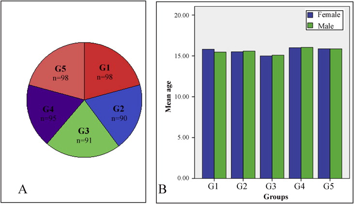
A sample of 374 adolescent patients (191 girls, 183 boys) with agenesis of at least 1 third molar was randomly selected from the files of orthodontic patients treated at the Department of Orthodontics at Karadeniz Technical University in Trabzon, Turkey. All patients were between 13 and 17 years old, had not received previous orthodontic treatment, and had no congenital anomalies. The patients were also checked to confirm that they had not undergone surgical removal or extraction of any of their third molars. Third molars were classified as developmentally missing when there was no evidence in the records that they had been extracted, or when there was no sign of tooth crown mineralization or crypt formation with radiolucency on panoramic radiographs. The patients were divided into 4 groups according to their third-molar agenesis patterns: group 1, agenesis of 4 third molars (98 patients); group 2, agenesis of 3 third molars (90 patients); group 3, agenesis of 2 third molars (91 patients); and group 4, agenesis of 1 third molar (95 patients). In addition, a control group (group 5) of 98 patients (51 girls, 47 boys) without third-molar agenesis was randomly selected from the patient archives to compare the distribution of the associated dental anomalies. The Figure shows the distribution and the mean ages of the patients in this study. Panoramic radiographs and cast models were used to determine the associated dental anomalies. The following dental anomalies were assessed.
Hypodontia: the absence of 1 to 5 teeth, excluding the third molar.
Hyperdontia: an excessive number of teeth in relation to the normal dental formula.
Impacted tooth: a permanent tooth with delayed eruption is an unerupted tooth whose root is developed in excess of this length and whose spontaneous eruption can be expected; a tooth that is not expected to erupt in a reasonable time, in these circumstances, is an impacted tooth.
Dilaceration: a deviation or bend in the linear relationship of a tooth crown to its root.
Peg-shaped maxillary lateral incisor: an incisor with a maximum mesiodistal crown diameter that is smaller compared with the same dimension of the opposing mandibular lateral incisor in the same patient.
Ectopic eruption: a tooth that, as a result of a disturbance in the eruption path, comes into contact apically to the prominence on the distal and mesial surfaces of the adjacent tooth, thereby locking the tooth.
Transposition: the positional interchange of 2 adjacent teeth, or a tooth’s development or eruption in a position normally occupied by a nonadjacent tooth.
Transmigrant canines: migration of a canine across the midline, regardless of the distance.
An x-ray technician performed all the radiographs using an orthopantomography device (Planmeca Proline CC 2002, Helsinki, Finland; 60-80 kVp, 8-10 mA, 12.8-second exposure time) with a magnification factor of 1.2. One investigator (M.C.) examined the dental anomalies.
Statistical analysis
To determine whether there were any methodologic errors, approximately 10% of the subjects (with or without third-molar agenesis) were randomly selected and evaluated by another researcher (M.B.) 4 weeks after the initial survey. There was 100% agreement between investigators. The Pearson chi-square and Fisher exact tests were used to determine the differences in the distribution of the associated dental anomalies between the groups and sexes. The Statistical Package for Social Sciences software (version 12.0; SPSS, Chicago, Ill) was used, with the significance level at P <0.05.
Stay updated, free dental videos. Join our Telegram channel
VIDEdental - Online dental courses


