The advances in the field of materials as they relate to orthodontics can be divided into the actual evolution of materials applied to daily practice and the changes in research methods to study the performance and the biologic properties of the materials. Although it is evident that new materials have saturated the market during the past century, the basic concepts of attaching one appliance to the enamel to use as a grip and inserting wires into that to control the spatial orientation of a tooth are identical to the original concepts. In contrast to that, the numbers of treatises about those subjects and the complexity of instrumentation and analytic tools used in published research have advanced tremendously and at a frenetic pace. This highly specialized pattern of research may effectively raise boundaries across research areas, since the complexity of the issues allows researchers to comprehend the content of journal articles in a narrow spectrum of disciplines. The purposes of this article were to review the advances in the research methods for investigating the various properties of orthodontic materials and to assist the reader in navigating this topic. A synopsis of the materials is also provided, listing future applications that already exist at the experimental stage or are yet unavailable but with the relevant technology already presented in broader scientific disciplines.
Highlights
- •
Materials used in daily orthodontic practices have evolved.
- •
Research methods to study the materials and their biologic properties have changed.
- •
This article reviews advances in research methods.
- •
A synopsis of the future of materials and applications is also presented.

The application of dental materials science in orthodontics coincides with the use of gold wire alloys by Edward Angle; the father of the specialty might not have imagined the impact that materials would have in current orthodontic practice. As the field progressed and grew to the dimensions of a specialty, the principles and mechanics of materials, which are typically taught in the first year of an undergraduate engineering curriculum, were incorporated in postgraduate orthodontic curricula along with accompanying elements of materials.
In the United States particularly, the disciplines of orthodontic mechanics and materials science received further emphasis, possibly because dental graduates entering orthodontic programs often had a bachelor’s degree in natural or engineering sciences, thus allowing for the cultivation and growth of materials research and bringing a new perspective to traditional and empirically taught concepts of the topic.
The advances in the field of materials as they relate to orthodontics can be divided into (1) the actual evolution of materials applied to daily practice and (2) the changes in research methods to study the performance and the biologic properties of the materials.
With respect to the first topic, although it is profoundly evident that new materials have saturated the market during the time period examined, the basic concepts of attaching one appliance to the enamel to use as a grip and inserting wires into that to control the spatial orientation of a tooth are identical to the original concepts. Of course, there have been different modes of attaching this “handle” to the tooth structure, along with many bracket materials and designs (lingual, self-ligating), and more alloy selection options are available, but the foregoing changes are within the path of the original concept.
Although aligners have revolutionized the conventional appliance configuration and constitute a new means of tooth movement, they can also be seen as representing a new version of the removable appliance tooth movement pattern that was popular in the middle of the previous century. Collectively, orthodontists practicing in the Angle era could easily have adapted, with only a few hours in a briefing seminar, to the conditions of the specialty 100 years later.
Nevertheless, the number of treatises about these subjects and the complexity of the instrumentation and analytic tools used in the published research have advanced tremendously and at a frenetic pace. For example, the engineering approaches in materials science and mechanics, the methods used to study the biologic mechanisms of tooth movement, and the data-analysis attributes of clinical trials require a strong background in the respective sciences involved; this makes it almost unattainable even for contemporary orthodontists to follow the developments across the various fields. This highly specialized pattern of research may effectively raise boundaries across research areas, in the sense that the complexity of issues allows researchers to comprehend the content of journal articles in a narrow spectrum of disciplines. This has been highlighted by studies investigating the characteristics of orthodontic publications, which showed a significantly higher frequency of multiauthor teams with affiliations from different scientific disciplines within a decade.
The purposes of this article are to review the advances in the research methods for investigating various properties of orthodontic applications of materials and to assist the reader in navigating this topic. A synopsis of the materials is also provided, listing future applications that already exist at the experimental stage or are yet unavailable but for which the relevant technology is already presented in broader scientific disciplines.
Evolution of orthodontic materials research approaches: A list of paradigms
The next paragraphs describe briefly the shift that has occurred in the past decades in the approaches to resolving several issues related to orthodontic materials and their applications. The subheadings lead the reader from the first steps of intervention beginning with bonding and covering appliances and their properties. This section is complemented with an overview of in-vivo aging studies or retrieval analyses, which constitute the greatest breakthrough in the area of simulation of the clinical environment.
Bonding
Roughness of enamel
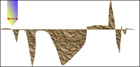
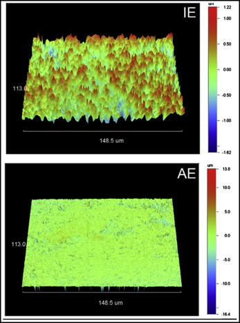
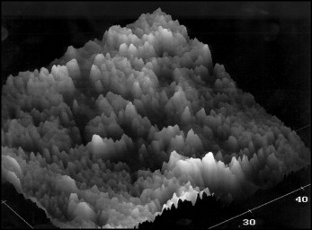
The alterations in the composition, topography, and roughness of enamel have been topics of research because of concern about potential irreversible changes caused by orthodontic bonding. The traditional approach to assessing roughness has been to use images of enamel (initially microscopic images and later scanning electron images) to quasi-quantify the effect of bonding on enamel appearance; this method is hampered by an apparent lack of sensitivity because the roughness variations of the surfaces can only be approximated. Later, a stylus-type of profilometric analysis of the surface of the substrate before and after bonding was used to investigate the increase in various roughness parameters, which are used to describe the variations of the surface (peaks, valleys, and so on). As shown in Figure 1 , the conventional approach with profilometry suffers from various limitations related mostly to the dimensions of the recording stylus; in the case of deep, narrow valleys, with dimensions smaller than the order of magnitude of the stylus, this method does not provide an accurate account of the extent of alterations ( Fig 1 ). The use of 3-dimensional optical profilometry bypasses this obstacle ( Fig 2 ), as does atomic force microscopy ( Fig 3 ), which, however, is essential for studying surface roughness at the nanoscale level, having a resolution far exceeding that of other stylus and optical-based methods, and which is somehow not applicable to large surface variations such as those expected after bonding. In the past 2 decades, we have come to understand far more on the topic than we have ever had, and this is largely due to the involvement of resources, material and human, from various scientific disciplines.
Polymerization efficiency (degree of C=C conversion)
Polymerization efficiency, also termed degree of cure or conversion, is often mistakenly noted as “degree of polymerization”; the latter represents the number of repetitions of the molecule of a monomer to become a polymer. The degree of cure is considered a key property for all polymers. In dental composite resins, this variable has been found to modulate the physical, mechanical, and biologic properties of the material, since a poor polymer network is susceptible to the release of biologically reactive substances (monomers and additives), predisposes to water absorption and swelling and hydrolytic degradation, and is associated with reduced mechanical properties. The latter had given rise to approaches that focused on the investigation of the phenomenon through mechanical testing with the hypothesis that since a higher degree of conversion is associated with an improved mechanical-properties profile of the material, evidence on the polymerization efficiency of the polymer could be extrapolated by measuring the response to various tests. However, the kinetics of the degree of cure and the mechanical properties at the time were not explored, and this resulted in erroneously assigning higher degrees of cure in materials that had better mechanical properties. Since the 1970s, through the pioneering work of Ruyter and Györösi, the degree of cure of orthodontic (and broader dental) resinous adhesives has been investigated with actual measuring of the extent of unreacted methacrylate groups, thereby allowing the percentage of converted double bonds (polymerization) to be accurately estimated on the surface of the materials.
Bond strength
Bond strength tests constitute much of the bonding-related literature, with more than 700 publications listed in the PubMed database during the last decade. Aspects of test standardization related to the direction of loads, morphologic variations of tooth crowns, source and age of extracted teeth planned to be used for testing, and maintenance of teeth in solutions have been analyzed, and the shortcomings of these preliminary tests have been demonstrated. The lack of aging of materials and the associated questionable clinical relevance of these tests has shifted the interest to more meaningful research, which provides an appraisal of the performance of the material complex (adhesive-bracket) in conditions identical to the actual clinical situation. Therefore, failure rate studies adopting a prospective study protocol or, even better, a randomized controlled trial design have gradually been appearing more often in the literature because of their direct clinical extrapolations. Nonetheless, the generalizability of the results of prospective or randomized controlled studies, albeit much wider than that of studies of bond strength, depends on the operators; thus, the results should not be interpreted without caution.
Prevention of demineralization
The attachment of orthodontic appliances to the enamel surface and their prolonged presence in the oral environment has been associated with the development of some unfavorable sequelae in the form of demineralization of hard dental tissues. Fluoride is the most potent cariostatic agent available that can prevent lesions, and dental hygiene plans have this as a central concept with the use of many materials at different stages such as bonding (glass ionomer cements) and during treatment (gels, rinsing solutions, and varnishes). For some applications such as glass ionomer cements or fluoride-releasing resinous adhesives, the long-term cariostatic effect of fluoride-releasing adhesives has not been sufficiently established, since most of the fluoride is released within the first few days or weeks. Therefore, topical fluoride in the form of solutions, varnishes, or gels has been an integral part of hygiene. A recent revolutionary approach targets the treatment of enamel after the formation of white spots; until now, this was limited to interventional restorations. Specifically, peptides such as a statherin-like peptide have been found to reduce the rate of hydroxyapatite demineralization in caries-simulating solutions by about 50%; research efforts have also focused on salivary proteins, which can bind to hydroxyapatite surfaces and form a selectively permeable pellicle.
Appliances
Corrosion
Corrosion of the orthodontic bracket-archwire complex has received attention after the corrosion products of the bracket base were shown to be diffused into the adhesive. The complexity of the materials and interfaces involved contributes to the development of corrosion in various elements of the appliance. For example, the bracket in its traditional configuration is composed of 2 phases: a low modulus of elasticity stainless steel alloy for the manufacturing of the base, which presumably allows for easy debonding after treatment, and a high-modulus steel alloy for the wings, which minimize deformation caused by the engagement of the wire and transfer the stresses from the activated archwire or the prescribed bracket slot to the tooth. These 2 alloys are joined by brazing alloys of nickel, silver, or gold; as a result, a galvanic corrosion is formed. In addition, the engagement of the wire into the slot with stainless steel ligatures formulates an environment in which many forms of corrosion can develop. For further information, the reader is referred to a thorough yet simplified review on the topic that lists all potential forms of corrosion and their mechanisms.
The advances in the field of orthodontic corrosion are largely due to the work of engineers, who laid the fundamentals for understanding the mechanisms of this phenomenon. The initial methods of simply examining the surface under incident light (low magnification microscopy) or of weighing the material before and after its exposure to the effector were replaced by research methods assessing the galvanic potential by measuring the possible differences. Studies in the field identified that the potential differences were positive, indicating that archwires were consistently the cathode, and brackets were the anode of the galvanic cell. Concurrently, imaging techniques advanced, and scanning electron microscopy in conjunction with energy dispersive analysis has contributed information on the topography, morphology, and elemental analysis of the areas of interest of the alloys.
Forces and moments of wire-bracket combinations in various setups
For the forces and moments of wire-bracket combinations in various setups, the traditional approach of most articles published in the early stages of research in orthodontic materials consisted of bench tests with simplified laboratory mechanical configurations, which in most cases were limited to 3-point bending and cantilever tests. Tensile tests for the extrapolation of the modulus of the material were also often used. An effort to simulate the orthodontic clinical analog had to overcome 2 basic obstacles: the complex mechanical profile of multiple brackets bonded to teeth in an arch form configuration, with wires engaged into the slots and ligated with elastomeric or stainless steel ligatures; and the aging of the materials in the intraoral environment and the effects on their properties. Although the latter was not resolved until recently, the first difficulty was overcome in a series of approaches.
1. Theoretical analysis with finite element analysis software was introduced in orthodontics from related sciences as a valid tool to predict the forces and moments developed during the engagement of wires as well as for the insertion of miniscrews and orthodontic implants.
2. Furthermore, experimental instrumentation included advanced analytic tools such as x-ray diffraction and differential scanning calorimetry, which, although they did not provide a direct result on the actual mechanical profile of the archwires, assisted essentially in explaining the performance of materials in mechanical tests and improved our understanding of the mechanisms underlying the load-deformation curve of these wires.
3. Concurrently, new complex mechanical testing configurations were proposed to simulate a bonded arch with various clinical malocclusion scenarios and a broad range of bracket and wire types. These in-vitro constructed analogs of clinical situations provided information that may be considered to be the closest to the intraoral situation if the effect of aging of the materials is excluded; that was a decisive step in obtaining data on the development of forces and moments in the 3 planes of space during the engagement of wires in brackets.
4. More recently, brackets with integrated strain gauge-type sensors were proposed; these are the most advanced appliances since the onset of our specialty. These can provide actual force ranges in either real time (in the case of bulky brackets, which carry a power source) or a retrospective fashion, which allows the manufacturing of slim forms of appliances.
Effect of intraoral conditions on materials
The area of the effect of intraoral conditions on materials has undergone extensive changes, to the point that it is a new field of research. Initially, the materials were subjected to aging by immersing them in various liquids (water, saline solution, artificial saliva, acidic liquids) for various times and then performing tests (mechanical properties, structural investigations, compositional alterations) to identify the effects of aging. Modern research relies on retrieval analysis, which was already a routine approach in the biomedical literature in the 1970s. Retrieval analysis goes beyond the scope of this article, and the interested reader is referred to an article on this topic.
After the 1990s, there was a specific interest in the investigation of the material performance through clinical trials as opposed to the extrapolation of information on intraoral behavior by in-vitro tests or simulations. This approach has provided information that rejected assumptions on various topics such as self-ligating brackets, the effectiveness of nickel-titanium wires, or the failure rates of brackets; further information on this topic can be found in this issue of the Journal .
Side effects of materials
In this field, the release of substances (ions from alloys and monomers, degradation by-products, and additives from polymers) and the biologic properties of materials are included.
Release of substances from orthodontic materials
In the early stages, efforts to study the release of substances from orthodontic materials consisted of measuring in vitro and with primitive techniques (weight, morphology) the ions or monomers released in the immersion media. Later, the same method was complemented with the introduction of instrument analysis such as atomic emission or absorption spectroscopy for metals. High-performance liquid chromatography and gas chromatography–mass spectroscopy were also used for the qualitative and quantitative analysis of immersion media, saliva, blood, or urine with respect to the concentrations of polymer by-products.
Biological properties of materials
With the establishment of a reliable method for the identification of released substances, the necessity for assessing the potential biologic reactivity of the materials emerged. The earliest methods were usually confined to adding various concentrations of effectors hypothesized to be released from orthodontic materials in cultures of immortal cell lines and observing the inhibition zone, which corresponded to the lack of levels of concentration causing cell death.
Later, more advanced techniques allowed for the assessment of actions at subtoxic concentrations to observe the effect on the physiology of a cell (or an animal) by using markers of oxidative stress or metabolic function and in 3-dimensional cultures. Then, with the availability of data on the quantities of species released, these levels of effectors can be added to human gingival or periodontal ligament fibroblasts to determine the corresponding mode of action at doses documented in vivo and to identify the potential effects on human cells.
Stay updated, free dental videos. Join our Telegram channel
VIDEdental - Online dental courses


