History of the Procedure
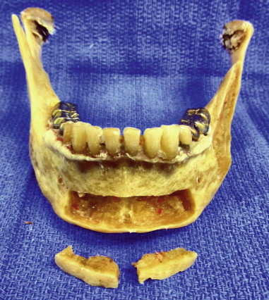
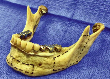
Intraoral bone graft techniques have been commonly used to repair maxillofacial bony defects. Historically, they have been used as dorsal nasal struts to repair orbital floor defects, augment alveolar ridges, repair clefts, and even for cosmetic augmentation in the paranasal region. The body/ramus and symphysis grafts are composed of autogenous intramembranous bone, which is easily accessible and therefore leaves no cutaneous scar ( Figures 126-1 and 126-2 ). Minimal morbidity is associated with these grafts, and they can easily be performed as in-office procedures, thereby decreasing the overall cost.
Indications for the Use of the Procedure
Harvest site selection depends on a thorough understanding of the quality and quantity of bone needed for the intended reconstruction. One graft may be selected over another, for example, if a higher content of cortical bone is desired. A symphysis graft offers a robust quantity of cancellous bone in addition to cortical bone. In contrast, a body/ramus graft provides a large quantity of bone and is an excellent source of dense cortical bone; however, only minimal cancellous bone is available.
The reported amounts of bone harvestable from intraoral sites vary throughout the literature, especially regarding total volume. The largest study to date performed on 59 cadavers looked at the surface area and volume harvestable from various intraoral sites ( Table 126-1 ).
| Donor Sites | Surface Area of Corticocancellous Graft (mm 2 ) | Volume of Corticocancellous Graft (mL) |
|---|---|---|
| Symphysis | 358.9 mm 2 | 1.15 mL |
| Body/ramus | 855.6 mm 2 | 2.02 mL |
Technique: Symphysis Harvest
Step 1:
Anesthetic
Inject local anesthesia with vasoconstrictor from the second premolar to the second premolar 7 minutes prior to incision.
Step 2:
Incision
Two incision designs are acceptable: sulcular and vestibular. The vestibular incision has a higher incidence of postoperative pain, scarification, dehiscence, decrease in vestibular length, and possible chin ptosis if the mentalis muscle is inadequately repaired. However, it is a viable option if the periodontal health of the mandibular incisors is poor. This is due to increased gingival recession that can occur with a sulcular incision in this patient population.
Technique: Vestibular Incision
Double skin hooks are placed on the lower lip to provide gentle retraction and allow adequate visualization of the anterior symphysis. Using a #15 blade or bovie electrocautery, the surgeon makes a curvilinear incision extending from the first premolar to the first premolar. Anterior to the canine region the vestibular incision should be 10 to 15 mm from the mucogingival junction; however, in the premolar region the incision should be only 5 to 10 mm from the mucogingival junction to avoid the mental nerve branches.
The incision is carried through mucosa only, to avoid the mental nerve branches. Avoiding the mental nerve branches, the surgeon incises the mentalis muscle using a curved Mayo at a 45-degree angle to the mandible, thereby simultaneously avoiding perforation through the skin and allowing myomucosal cuff preservation. Once at the periosteum, the surgeon incises using a #15 blade or Bovie electrocautery.
Step 1:
Dissection
A full thickness mucoperiosteal flap is developed using a periosteal elevator in all directions until the mental nerve foramen is identified bilaterally and the inferior border of the mandible exposed.
Step 2:
Stay updated, free dental videos. Join our Telegram channel
VIDEdental - Online dental courses


