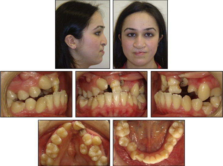
The comprehensive treatment of a patient with cleft lip and palate requires an interdisciplinary approach for functional and esthetic outcomes. A 20-year-old woman with bilateral cleft lip and palate had a chief complaint of unesthetic appearance of her teeth and the presence of oronasal fistulae. Her clinical and radiographic evaluation showed a dolichofacial growth pattern, a Class II skeletal relationship with retroclined maxillary central incisors, 5 mm of negative overjet, maxillary constriction, maxillary and mandibular crowding, congenitally missing maxillary right incisors and left lateral incisor, and a transposed maxillary left canine. Her treatment plan included the extraction of 3 premolars, maxillary expansion, segmental maxillary osteotomy, repair of the oronasal fistulae, rhinoplasty, periodontal surgery, and prosthodontic rehabilitation. To obtain a better occlusion and reduce the dimensions of the fistulae, orthognathic surgery comprising linear and rotational movements of the maxillary segments (premaxilla, right and left maxillary alveolar segments) in all 3 axes was planned by performing 3-dimensional virtual surgery on 3-dimensional computerized tomography. At the end of the interdisciplinary treatment, a functional occlusion, a harmonious profile, and patient satisfaction were achieved. Posttreatment records after 1 year showed stable results.
Cleft lip and palate (CLP) is a congenital deformity that is associated with maxillary sagittal, transversal, and vertical discrepancies. In addition to skeletal discrepancies, this deformity is often accompanied by dental abnormalities, such as hypodontia, hyperdontia, and transpositions. Hypodontia, especially the absence of the maxillary lateral incisors, is the most prevalent. The combination of the skeletal and dental malocclusion with the soft-tissue and hard-tissue deformities or deficiencies complicates the treatment of CLP and requires interdisciplinary approaches to obtain functional and esthetic outcomes.
In recent years, 3-dimensional (3D) virtual planning of orthognathic surgery has begun to be used in clinical practice. Computer-aided surgical simulation enables doctors to perform osteotomies, reposition the osteotomized bony structures, control intercuspation, control interferences between osteotomized bony structures, evaluate difficulties before surgery, and simulate the postoperative results on the hard and soft tissues in 3 dimensions. The challenging treatment of CLP can benefit from 3D surgical planning because of the complex characteristics of the malocclusion and the patient’s unique and individual requirements. In patients with alveolar clefts in whom integrity of the alveolar segments was not achieved, treatment becomes multipiece maxillary segmentation after LeFort I osteotomy. When multipiece maxillary segmentation is used, it is not possible to describe the intraoperative movements of each maxillary segment with conventional surgical planning. Computer-aided surgical simulations have advantages, especially for these patients.
The aim of this case report was to present the interdisciplinary treatment of a 20-year-old woman with bilateral CLP and congenitally missing and transposed teeth.
Diagnosis and etiology
A 20-year-old woman with operated nonsyndromic bilateral CLP was referred to the orthodontic clinic of Yeditepe University in Istanbul, Turkey. Her chief complaints were the unesthetic appearance of her teeth and the presence of oronasal fistulae. She had received primary lip repair and palatoplasty in the first year of life and did not undergo bone grafting. Her extraoral examination showed a slight asymmetry at the eye level but no apparent mandibular asymmetry. She had nasal deviation, widening of alar bases, and loss of columellar projection. Her retruded upper lip was improperly repaired and scarred. Her lower lip was protruded. Her V-shaped maxillary arch was severely constricted. The premaxillary segment was mobile. Her intraoral photographs showed anterior and posterior bilateral crossbites, severe maxillary and mandibular crowding, a deep curve of Spee, Angle Class II molar relationships, a palatally displaced and rotated left second premolar, a transposed maxillary left canine, and a hypomineralized maxillary central incisor. The maxillary left deciduous canine had extensive caries. Bilateral alveolar fistulae were present ( Figs 1 and 2 ).


