6
IMPACTED TEETH
Impacted teeth are a common problem. Orthodontists and oral and maxillofacial surgeons spend a lot of time assessing tooth position and eruption patterns and managing patients referred from general dentists who have usually seen these impactions on intraoral or panoramic radiographs. Permanent canines erupting abnormally are common, as are horizontally impacted mandibular third molars. Even supernumerary teeth are a common enough anomaly to require additional radiographic assessment. Cone beam volumetric imaging (CBVI) is the most appropriate way to perform this assessment for preoperative planning and orthodontic management. It is likely that CBVI will become the standard of care for the assessment of all impactions in the near future.
FIG 6-1
MAXILLARY CANINE AND MANDIBULAR THIRD MOLAR
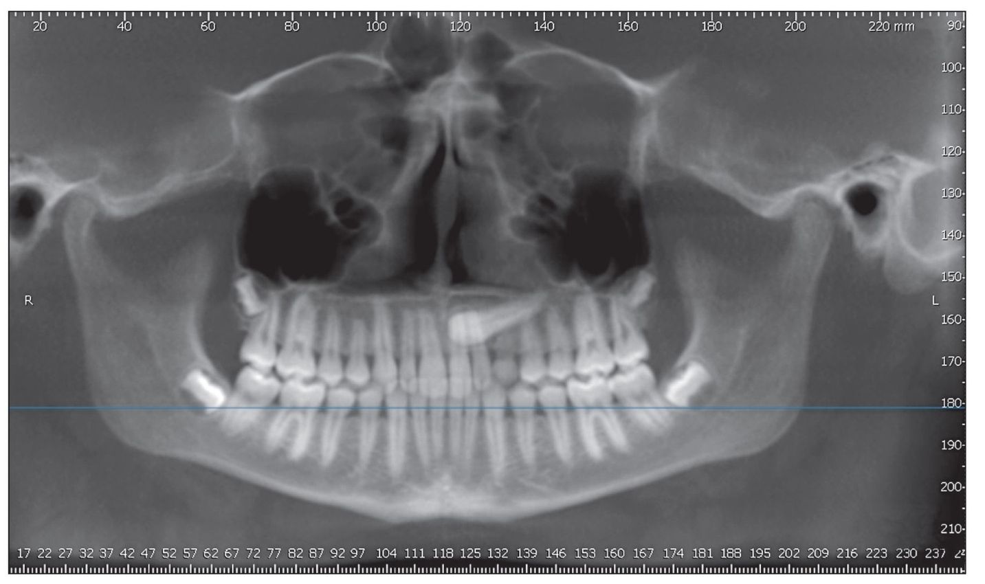
Fig 6-1a A panoramic image, reconstructed from the cone beam data volume, represents the type of image that would serve for the initial assessment of the missing canine.There is no way to determine the correct orientation (facial or palatal position) from this panoramic image.The primary canine is retained.The permanent canine is impacted horizontally.y.
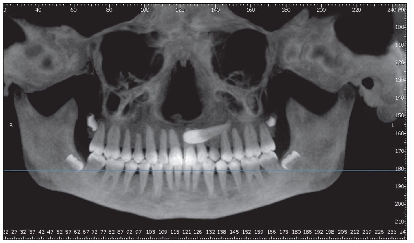
Fig 6-1b The same image as Fig 6-1a, using a maximum intensity projection view. The canine appears to be anterior to the central and lateral incisors.
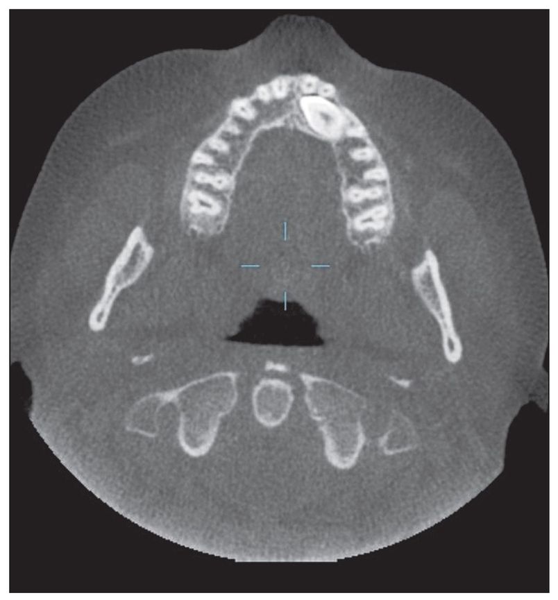
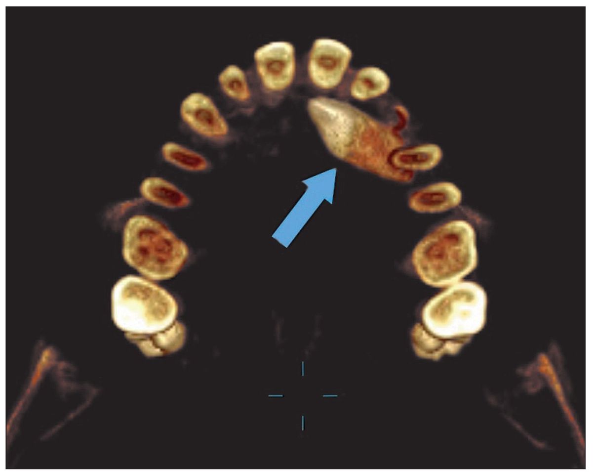
Fig 6-1c The cone beam multiplanar reconstructed (MPR) axial image reveals the correct position of this impacted canine. It is posterior to the central and lateral incisors and the retained primary canine.
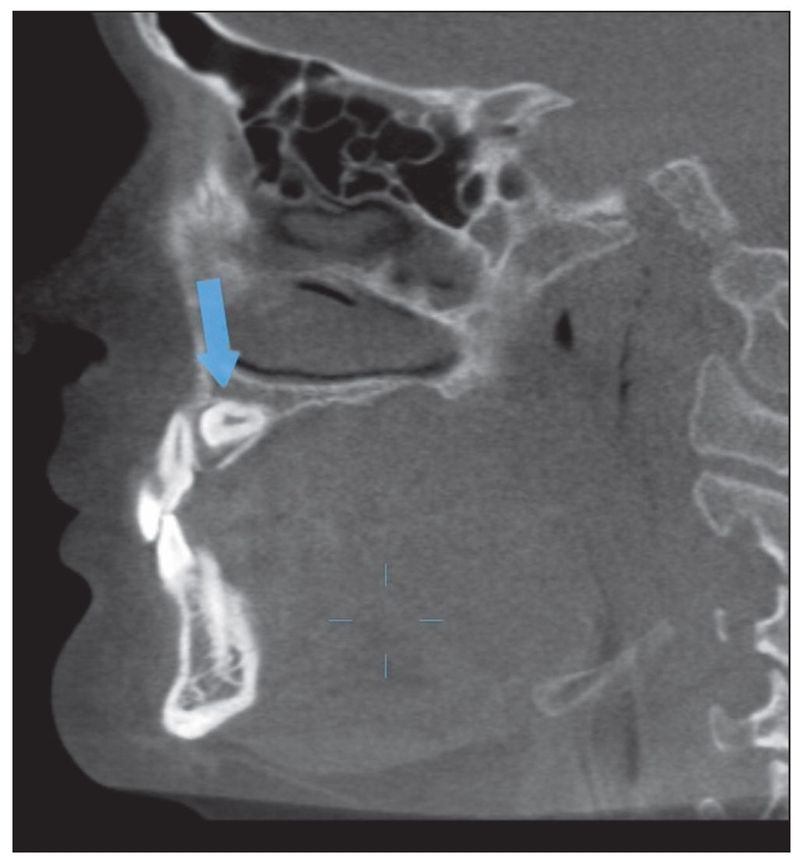
Fig 6-1d The cone beam MPR sagittal image reveals the position of this impacted canine (arrow) relative to the left lateral incisor.
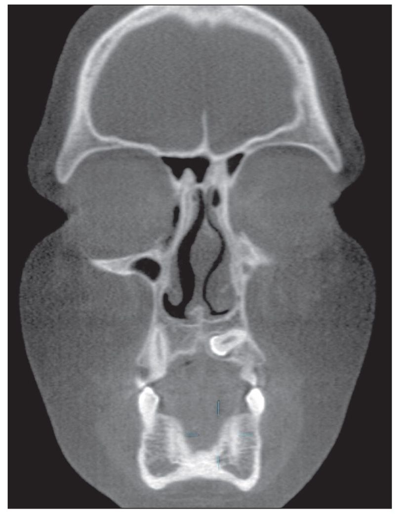
Fig 6-1e The cone beam MPR coronal image reveals the position of this impacted canine as posterior to the central and lateral incisors.
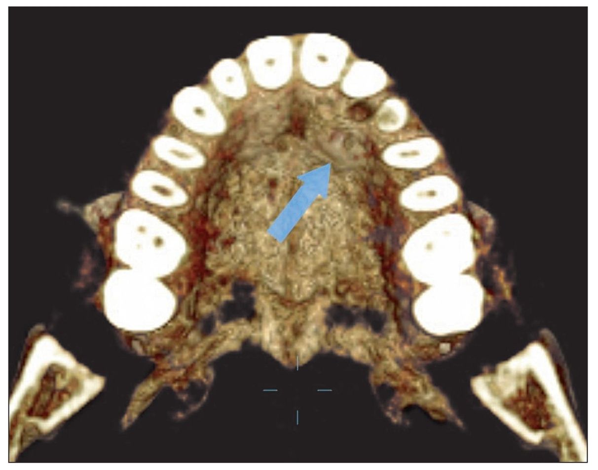
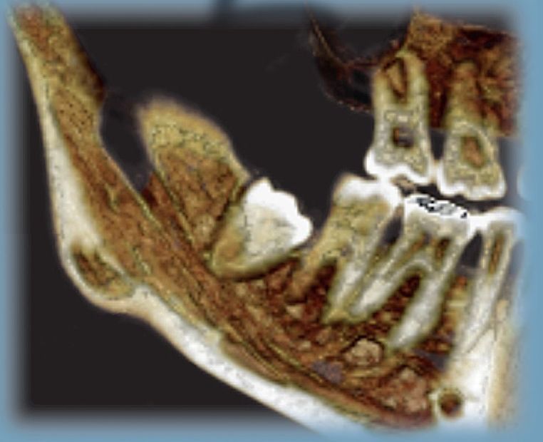
Fig 6-1f A 3-D color reconstruction shows the palatal elevation caused by the impacted canine (arrow).
Stay updated, free dental videos. Join our Telegram channel
VIDEdental - Online dental courses


