Introduction
Our objective was to measure the impact on perceived root resorption based on the amount of anteroposterior incisal inclination as determined in vitro from conventional panoramic radiography.
Methods
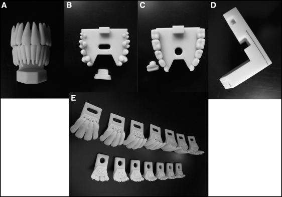
A rapid prototyping model was created to mimic different maxillary and mandibular incisal anteroposterior inclinations. Two titanium beads were placed on the incisors at the apical and incisal edges. Panoramic radiographs were obtained, with the incisors changing relative inclination by 10° increments. The length was measured from the midpoint of the bead on the incisal edge to the midpoint of the bead on the apical edge. By using a length of wire of known size, this value was compared in all images to correct for image magnification.
Results
Changes to mandibular incisor anteroposterior inclinations, as either a theoretical proclination or retroclination, resulted in an increase of “apparent” root resorption on a panoramic radiograph. When the maxillary incisors were significantly and severely retroclined, they appeared larger than expected. When the maxillary incisors were mildly retroclined, the length was roughly similar to the theoretical model. When the maxillary incisors were mildly proclined, they appeared shorter than expected.
Conclusions
The foreshortening or forelengthening of incisor root lengths because of incisor inclination vs root resorption cannot be reliably evaluated from panoramic images. The proposed theoretical model helps to understand the direction of the changes produced by the magnification factor. More severe scenarios where either the maxillary or the mandibular teeth are outside the focal trough have not been fully evaluated. The clinical impact of these changes is likely to be questionable.
Highlights
- •
Incisor external apical root resorption was assessed on panoramic radiographs.
- •
As mandibular anteroposterior incisor angulation changed, teeth appeared shorter.
- •
Shorter teeth were associated with increased external apical root resorption.
- •
Actual impact of root foreshortening caused by imaging limitations is questionable.
A thorough radiographic examination is part of the diagnostic process in orthodontics. Lateral cephalometric and panoramic films are routinely ordered as the primary pretreatment radiographs.
Clinicians often assess whether root resorptive changes have occurred in teeth undergoing orthodontic treatment with panoramic radiographs. Panoramic radiographs are not likely to accurately differentiate a small amount of external root resorption from normal root length, so in cases of suspected orthodontically induced external root resorption, further imaging with periapical radiographs may be prescribed. Assessment of root length using 2-dimensional radiographs must be done with caution because the radiographic image is often affected by distortions in root angulation and magnification.
A panoramic film displays both the maxillary and the mandibular arches as well as the supporting structures in 1 convenient image. The quality of the generated image on a panoramic radiograph relies on a focal trough that is similar in shape to a dental arch and resembles a 3-dimensional horseshoe. To obtain the best image on the panoramic image, the structures of interest need to be within the focal trough. Sometimes, especially when a patient has significant skeletal anteroposterior discrepancies, important structures such as the maxillary and mandibular incisors can end up outside the focal trough, resulting in distorted or obscured incisors in the radiographic image.
In orthodontic camouflage of mild or moderate Class II patients, the mandibular incisors may become excessively proclined, leading to an apparent foreshortening of the roots as seen in the radiographic image. By developing an appreciation for the predicted amount of foreshortening that can occur because of incisor angulation, clinicians may be better able to recognize true root resorption vs apparent foreshortening.
Therefore, the objective of this study was to measure the effects of anteroposterior incisal inclination on the resulting perceived incisor length produced on conventional panoramic images from a rapid prototyping model.
Material and methods
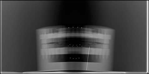
Two titanium beads (Abbott Ball, West Hartford, Conn) were placed on a rapid prototyping model of each maxillary and mandibular incisor at their apical and incisal edges. The maxillary and mandibular arches were fit into a special holding device where maxillary and mandibular incisor segments of various angulations were fitted to their respective arches using a lock and key type of jig ( Fig 1 ). Panoramic imaging ( Fig 2 ) was acquired with the same machine (Orthophos XG; Sirona Dental Systems, Long Island City, NY), and the produced image was stored in software (Dolphin Imaging & Management Solutions, Chatsworth, Calif). The panoramic images were then hand-traced and measured with a digital caliper. The length was measured from the midpoint of the bead on the incisal edge to the midpoint of the bead on the apical edge. This value was compared with the known length, measured from the incisal bead to the apical bead on the rapid prototyping model minus the diameter of 1 bead. By using a length of wire of known size, this value was compared in all images to correct for any image magnification.
To account for magnification, the length of the tooth was calculated as follows.
(actual length of calibration wire/measured length of calibration wire on radiograph) * length of tooth on radiograph
The theoretical lengths of the incisors from the rapid prototyping models were calculated after adjusting for the inclination of the incisors using the equation
theoretical length = sin (Θ) * measured length of incisors on the actual rapid prototyping models
Results
The reliability of the hand-tracing using the intraclass correlation coefficient (ICC) was excellent for the maxillary incisors and the mandibular right permanent lateral incisor. The ICC was fair to good for the remaining mandibular incisors ( Table I ).
| Incisor | ICC (95% CI) | Reliability |
|---|---|---|
| Maxillary right lateral | 0.978 (0.916-0.996) | Excellent |
| Maxillary right central | 0.957 (0.839-0.992) | Excellent |
| Maxillary left central | 0.947 (0.803-0.990) | Excellent |
| Maxillary left lateral | 0.966 (0.874-0.994) | Excellent |
| Mandibular right lateral | 0.870 (0.516-0.976) | Fair-good |
| Mandibular right central | 0.920 (0.703-0.985) | Fair-good |
| Mandibular left central | 0.862 (0.485-0.974) | Fair-good |
| Mandibular left lateral | 0.951 (0.819-0.995) | Excellent |
While the actual measurements are presented in Table II , the theoretical expected measurements are presented in Table III . The difference between both measurements is depicted in Table IV .
| Maxillary central incisor angulation (°) | Maxillary lateral incisor angulation (°) | Right lateral (mm) | Right central (mm) | Left central (mm) | Left lateral (mm) |
|---|---|---|---|---|---|
| 49.8 | 52.3 | 23.64 | 25.26 | 25.76 | 23.89 |
| 59.8 | 62.3 | 25.05 | 26.87 | 26.69 | 24.98 |
| 69.8 | 72.3 | 26.17 | 27.53 | 27.06 | 25.87 |
| 79.8 | 82.3 | 26.47 | 27.95 | 27.75 | 26.50 |
| 89.8 | 92.3 | 25.95 | 26.95 | 27.03 | 26.11 |
| 99.8 | 102.3 | 25.04 | 25.86 | 25.92 | 24.98 |
| 109.8 | 112.3 | 22.94 | 22.93 | 22.70 | 22.53 |
| Mandibular central incisor angulation (°) | Mandibular lateral incisor angulation (°) | Right lateral (mm) | Right central (mm) | Left central (mm) | Left lateral (mm) |
|---|---|---|---|---|---|
| 140.5 | 140.2 | 12.27 | 12.27 | 12.33 | 13.14 |
| 130.5 | 130.2 | 15.40 | 15.40 | 15.47 | 15.62 |
| 120.5 | 120.2 | 17.90 | 17.90 | 17.85 | 17.51 |
| 110.5 | 110.2 | 18.69 | 18.69 | 19.03 | 18.77 |
| 100.5 | 100.2 | 19.85 | 19.85 | 19.81 | 19.99 |
| 90.5 | 90.2 | 20.75 | 20.75 | 20.89 | 20.11 |
| 80.5 | 80.2 | 20.22 | 20.22 | 20.74 | 20.11 |
| Maxillary central incisor angulation (°) | Maxillary lateral incisor angulation (°) | Right lateral (mm) | Right central (mm) | Left central (mm) | Left lateral (mm) |
|---|---|---|---|---|---|
| 49.8 | 52.3 | 20.70 | 20.97 | 20.94 | 20.92 |
| 59.8 | 62.3 | 22.94 | 23.58 | 23.70 | 23.17 |
| 69.8 | 72.3 | 25.26 | 26.12 | 25.73 | 24.96 |
| 79.8 | 82.3 | 26.04 | 27.52 | 27.15 | 26.10 |
| 89.8 | 92.3 | 26.37 | 27.54 | 27.53 | 26.22 |
| 99.8 | 102.3 | 26.07 | 27.41 | 27.41 | 25.78 |
| 109.8 | 112.3 | 24.31 | 25.95 | 25.80 | 24.60 |
| Mandibular central incisor angulation (°) | Mandibular lateral incisor angulation (°) | Right lateral (mm) | Right central (mm) | Left lateral (mm) | Left lateral (mm) |
|---|---|---|---|---|---|
| 140.5 | 140.2 | 12.99 | 12.99 | 13.07 | 13.03 |
| 130.5 | 130.2 | 15.17 | 15.57 | 15.44 | 15.36 |
| 120.5 | 120.2 | 17.20 | 17.59 | 17.36 | 17.35 |
| 110.5 | 110.2 | 18.61 | 19.01 | 18.99 | 18.73 |
| 100.5 | 100.2 | 19.60 | 19.80 | 19.97 | 19.59 |
| 90.5 | 90.2 | 19.87 | 20.50 | 20.19 | 20.03 |
| 80.5 | 80.2 | 19.74 | 19.94 | 20.39 | 19.59 |
Stay updated, free dental videos. Join our Telegram channel
VIDEdental - Online dental courses


