Fig. 8.1
This is a burn victim identification case where the lower jaw was removed to facilitate the taking of radiographs. The anterior teeth are completely charred, however the remaining sockets can be useful in identification

Fig. 8.2
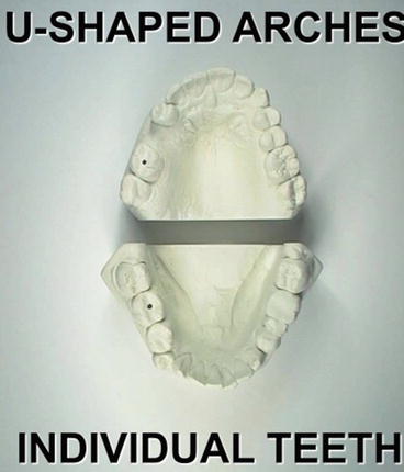
Indicates the criteria for recognizing a bitemark as human

Fig. 8.3
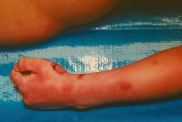
A bitemark on the left hand of a homicide victim. Numerous “defensive wounds” (bruises can be seen on her arm)

Fig. 8.4
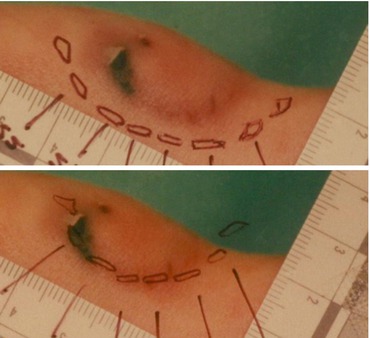
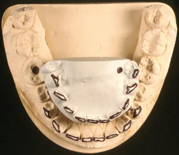
Impression of both the deceased victim and suspect were made and transparent overlays were created

Fig. 8.5
The victim in Slide 315 demonstrating the use of transparent overlays and the ABFO scales to compare the dentitions of both the suspect and the victim with the bitemark. In this particular case, it was determined that the victim bit herself during the perimortem period
With increasing use of dental cosmetic resin restorations, it is sometimes difficult to visually and radiographically scrutinize modern restorations in order to make needed comparisons [31]. Laboratory based scanning electron microscopy with energy dispersive X-ray spectroscopy (SEM/EDS) used in conjunction with the spectral library identification and classification explorer (SLICE) software can help identify resin material in an extracted tooth (Bush et al. 2008). Another resin identification method option used in the morgue or field is the portable X-ray fluorescence spectrometer (XRF) paired with a custom spectral library database to identify manufactured brands of resin dental materials ([4, 5]; Bush et al. 2008). Dentists should have the victim’s AM record noted with the brand names of restorative materials used so that information gathered by the SEM/EDS or XRF can help add to the certainty of identification ([4]; Senn and Stimson 2010). Still, AM and PM dental radiographic comparisons are the main odontological method used for identifications, but when traditional methods have been exhausted, SEM/EDS and XRF may be needed [4].
8.2.3 Cone-Beam Computed Tomography
Three-dimensional imaging in the form of cone-beam computed tomography (CBCT) is gaining popularity for clinical diagnostics and treatment planning in dental specialties. These highly detailed images become a permanent part of the patient’s dental record, and could serve as sources of AM information. Forensic odontologists can also use CBCT in order to obtain PM information, especially in cases when intraoral access and 2 dimensional (2D) dental radiography are not possible [14].
8.2.4 Computed Tomography, Magnetic Resonance Imaging and Surface Scanning
Enough detailed information from the deceased victim needs to be obtained in order to compare against AM data so that a positive identification can be confirmed. However, this is sometimes to achieve due to physical manipulation difficulty or religious objections to invasive means raised by the victim’s family [19]. In cases where PM dental information needs to be obtained, but is not possible through invasive surgical means, computed tomography (CT) is an option [19, 29]. This advanced three-dimensional (3D) digital radiography system yields detailed information while leaving the body intact. When available, mobile CT scanner vehicles equipped with distance reporting telecommunications could assist efforts at a MFI temporary morgue or onsite for crash accidents [29]. Comparisons of 2D dental radiographs are considered the gold standard for establishing dental unique identifiers, but CT images can be utilized as a complementary tool [3]. While sometimes criticized for its inability to give discriminatory information that is detailed enough for comparing dental restorations, it has been found to be valuable for age estimation in cases involving child victims [3].
When CT is combined with magnetic resonance imaging (MRI) and surface scanners, a non-invasive virtual autopsy makes it possible to obtain a large amount of PM information; including dental [11, 18]. An example of such a system is the Virtopsy project, which relies on a robotic system guided by three-dimensional surface scanning and software in order to collect autopsy information on fragile human remains without the risk of physical damage to the body [9, 28]. To begin, a digital SLR camera and optical surface scanner collect photogrammetry and triangulation imagery data that is wirelessly communicated to a software responsible for calculating millions of surface points on the skin, which navigate a robotic arm on how to perform the virtual autopsy task [28]. If the equipment is available during DVI events occurring in remote areas, the planning and commanding of the robotic arm can be carried out through wireless communications by experts who are off-site [9]. Virtual autopsy is a new and very advanced technique which is still being studied [19].
8.2.5 Radio Frequency Identification Tags
Using dental records to identify a person by unique characteristics found within the dentition is a common and well rehearsed activity among odontologists [26, 27]. However, identifying an edentulous person can be quite challenging due to the loss of unique odontological identifiers; making marked dentures invaluable for edentulous victims [20]. Methods for labeling oral prostheses ranges from surface notations in the form of handmade engravings or pen marks, to structural inclusion techniques such as thin papers, film or metal labels with a clear protective coating [8, 26]. These methods tend to be cost effective, simple to accomplish, and make identification easier than having no markings at all. However, they can have drawbacks in the form of esthetics, the potential for trapping bacterial plaque, and being susceptible to not holding up to the destructive nature of extreme heat, acids, or wear [8, 26]. Alternative methods that are quick and easy to access include electronic information storing microchips, barcodes, or radio frequency identification tags (RFID tag) that can be structurally included into the denture [7]. A major benefit of RFID tag use is its durability against harsh assaults such as extreme heat, freezing temperatures, wear, or acid exposures [26]. The wireless electronic communication data carrying tag contains a microchip with an antenna and is rated to have an unlimited lifespan [26, 27]. Identification information is accessed by a mobile handheld reader with an electromagnetic field emitting antenna to energize the transponder (tag). The tag gives the coded data to the reader, which translates the data into readable information [25]. Information stored on the tag can be changed over time as needed and is capable of holding contact information in addition to the name of the wearer.
8.2.6 Three-Dimensional Craniofacial Reconstruction
Forensic odontological and anthropological examinations of skeletonized remains can offer evidence of age, gender, and ethnicity so that a 3D craniofacial reconstruction can recreate an AM likeness [33, 34]. This is particularly important when there is no source of AM information for the unknown victim [6]. Advantages of 3D computerized reconstructions over traditional manual clay methods includes time efficiency, less subjective influences, characteristic variations can be applied for multiple renderings, and the digital image can be easily shared from a distance [17]. When the resulting image is released to the public, someone may recognize the victim and come forward to give authorities information to aid the investigation [6].
8.2.7 Software and Databases
Identifying a deceased victim relies heavily on comparisons of collected AM and PM dental information. Comparison software such as WinID3, Plass Data’s DVI System, and Unified Victim Identification System (UVIS) enables collected data to be electronically catalogued and filtered [14, 37]. Software algorithms automate comparisons based on dental patterns and other unique identifiers so that records can be ranked by numbers of matches, mismatches, and possible matches [2]. Prior to such software systems, odontologists spent a lot of time manually comparing multiples of AM records against each PM record of interest that was within their possession [14]. In addition to saving a lot of time, these software systems can be compared against databases for missing persons since they have a high probability of being a deceased victim odontologists will attempt to identify. Missing persons databases available for searches in multiple languages include UVIS Dental Identification Module (UVIS/UDIM) and NamUS (the National Missing and Unidentified Persons System) [36, 37]. These databases are accessible by the internet and can assist in the identification of unknown human remains [36, 37].
8.3 Bitemark Investigations
8.3.1 Bitemark Identification
Bitemarks are prevalent in cases where a victim has attempted to defend themselves from a perpetrator. A bite can be defined as a mark made by human or animal teeth on the skin of “living individuals, cadavers, or objects with relatively softened consistence” [13], p. 45. Bitemark analysis can be of assistance to the forensic team when attempting to understand if time has elapsed between the production of the mark and the examination. Analysis can also assist to determine if the marks occurred antemortem or postmortem, and in the case of multiple bites, the sequence of bite production can yield useful information [13] Fig. 8.2.
Bitemarks can be located anywhere on the body, however they are most often identified in areas of soft, fleshy tissues such as the face, stomach and buttocks [18]. When identifying bitemarks on a victim and dental records are not available, unusual oral characteristics gleaned from the teeth are used to narrow down clues based on the biter’s lifestyle habits such as wear patterns, chipping of teeth, dietary patterns, and socioeconomic strata [18]. In addition to unique conditions present, identifiers such as formation of eruption and dental aging based on cementum-formation and root -transparency level may assist in analysis of the data.
Stay updated, free dental videos. Join our Telegram channel

VIDEdental - Online dental courses


