Introduction
In this study, we evaluated root and alveolar bone development in unilateral osseous impacted immature maxillary central incisors by cone-beam computed tomography before and after closed-eruption treatment, in comparison with naturally erupted contralateral immature maxillary central incisors.
Methods and Results
The study included 30 patients, 20 boys and 10 girls, with a mean age of 8.44 ± 1.20 years (range, 6.5-11.2 years). After treatment, the root lengths of both the impacted maxillary central incisors (10.66 ± 2.10 mm) and the contralateral maxillary central incisors (11.04 ± 1.76 mm) were significantly greater than their pretreatment values (6.67 ± 1.94 and 9.02 ± 2.13 mm, respectively). The root canal widths of the incisors decreased significantly after treatment. From the posttreatment cone-beam computed tomography images, the ratio of exposed root length to total root length and the thickness of the alveolar bone at 1 mm under the alveolar crest and at the apex were calculated to evaluate alveolar bone development. Impacted immature maxillary central incisors differed significantly from contralateral immature maxillary central incisors in labial exposed root length, labial ratio to total root length, and lingual alveolar crest. Clinical crown height was higher (statistically but not clinically) for the impacted incisors (9.87 mm) than for the contralateral incisors (9.37 mm).
Conclusions
Impacted immature incisors grew to the same stage as did erupted contralateral incisors after closed-eruption treatment. Both incisor types had some alveolar bone loss, and thin alveolar bone surrounded the roots.
Highlights
- •
We evaluated root development in 30 patients after unilateral closed-eruption therapy.
- •
Alveolar bone development of osseous impacted maxillary central incisors was assessed.
- •
Impacted immature incisors grew similarly to contralateral incisors after treatment.
- •
Gingival margin positions differed between impacted and contralateral incisors.
- •
Periradicular osseous tissue reconstruction also differed between groups.
The maxillary central incisors are the most prominent teeth in the mouth, significantly affecting a child’s facial appearance, esthetics, pronunciation, mastication, and psychology. Although the canine is the most frequently impacted tooth in the anterior region (incidence, 1%-3% ), an impacted maxillary central incisor is the most conspicuous to parents.
Many studies have demonstrated that the closed-eruption technique is an effective method of treating impacted teeth. A strong positive relationship has been found between the necessary duration of this treatment and the patient’s age, and many researchers have reported that treatment begun in younger patients yields better results.
However, much of this research into the closed-eruption technique showed important problems that need to be solved. The patients selected in most studies included children, adolescents, and even adults. The root development of the impacted teeth was unclear. Most researchers studied only posttreatment examinations, with no comparison of pretreatment and posttreatment records. In most cases, the radiologic records used were periapical, panoramic, and cephalometric radiographs, which are not as accurate as cone-beam computed tomography (CBCT), especially on rotated and dilacerated teeth. In many patients, it was found that the impacted tooth roots were rotated before the treatment, and the roots of most impacted teeth were dilacerated. Thus, a convincing evaluation of the treatment efficacy was not possible.
The aim of our study was to evaluate the root and alveolar bone development of unilateral osseous impacted immature maxillary central incisors by CBCT before and after early closed-eruption technique. Naturally erupted contralateral immature maxillary central incisors were used for comparison.
Material and methods
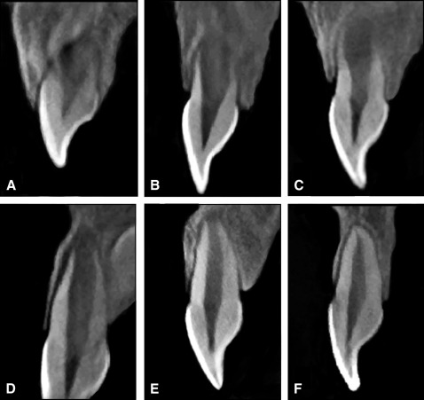
A total of 244 patients with impacted maxillary central incisors were consecutively treated by one operator (S.Z.) in the Departments of Pediatric and Preventive Dentistry at the Peking University School and Hospital of Stomatology. Of these, 30 patients (20 boys, 10 girls) were included in our study. These patients met the following criteria: (1) the coexistence of a unilateral osseous impacted maxillary central incisor, whose root was in stages 1 to 5 ( Fig 1 ) at the beginning of the closed-eruption treatment, with a contralateral maxillary central incisor (control) that had already erupted but had not necessarily reached the occlusal plane; (2) the closed-eruption treatment had been finished for about 1 to 3 years; (3) complete diagnostic and treatment notes were available; (4) pretreatment and posttreatment CBCT records were available; (5) there was no mechanical obstacle to eruption: eg, supernumerary teeth, tumors, odontoma, or cysts; (6) the patient had no systemic disease; and (7) the patient and parents cooperated with the treatment plan and provided informed consent. The exclusion criterion was an injury to the maxillary frontal area before our study finished.

A diagnosis of impaction was evaluated clinically and radiologically when one immature maxillary central incisor was absent from the dental arch after the expected eruption time and the contralateral incisor had erupted at least 6 months earlier, or the eruption orientation of one central incisor was not toward the center of the alveolar ridge (confirmed by radiologic examination).
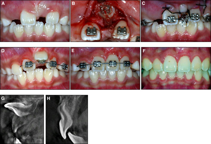
For the closed-eruption technique, a medical history was taken, and clinical and radiologic examinations (including CBCT) were conducted by a pediatric dentist (S.Z.) ( Fig 2 , A and G ). Impacted immature maxillary central incisors were treated by this same dentist with a combined surgical-orthodontic technique. The impacted incisors were exposed with a full-thickness mucoperiosteal flap, and a bracket was bonded to the teeth during surgery. This was tied to an orthodontic elastomeric chain (Clear Generation II power chain, closed space 639-0002; Ormco, Orange, Calif) for orthodontic traction. A 0.016 × 0.022-in rectangular stainless steel archwire was used to obtain adequate anchorage, with an omega-shaped bend applied to enhance the retention of the elastomeric chain between the bracket and the wire. The elastomeric chain was at its original length. The flap was then repositioned over the incision and sutured. The sutures were removed on postoperative day 7 ( Fig 2 , B and C ).

We cut off part of the elastomeric chain and reconnected it to the archwire to obtain 100 g of orthodontic traction force, measured with a testing machine (model 5969; Instron, Norwood, Mass), every 4 to 5 weeks. When the surface of the impacted incisor was exposed, a new bracket was bonded, and a 0.012-in round nickel-titanium archwire was used to guide the impacted central incisor toward the center of the alveolar ridge ( Fig 2 , D ). There were 19 patients who had insufficient space to accommodate the impacted incisors before treatment. Two had only 5.5 mm of space and needed orthodontic expansion before treatment of the impacted tooth, whereas in the other 17 patients the midline of their maxillary incisors declined for 1 to 2 mm; this could be corrected during traction of the impacted incisors. Thus, the patients could receive treatment without delay. When the extruded incisor was aligned in the dental arch, the 0.012-in round archwire would be replaced by a 0.016 × 0.022-in rectangular nickel-titanium archwire if the axis of the extruded incisor was inclined. When the impacted incisor was brought into good alignment with the adjacent teeth, the closed eruption was considered complete.
The retention period began when a 0.016 × 0.022-in rectangular stainless steel archwire was placed in the labial brackets of the central and lateral incisors ( Fig 2 , E ) as a fixed retaining appliance, without orthodontic force. Root and alveolar bone development was evaluated every 3 months by x-rays (periapical films).
Posttreatment CBCT images were obtained a minimum of 12 months later. If the retention period lasted more than 12 months, posttreatment CBCT was used after debonding; otherwise, CBCT images were taken at 12 months after the end of the treatment period ( Fig 2 , F and H ).
Patient charts were reviewed for the following information: age, sex, medical history, bonding date, surgical exposure date, debonding date, and posttreatment CBCT examination date. The duration of the orthodontic traction time was calculated as the time between the application of the power chain and good alignment of the impacted incisor in the dental arch. The duration of the retention time was calculated as the time between the alignment of the impacted incisor in the dental arch and the debonding date of the maxillary central incisors. The duration of the follow-up time was calculated as the time between the debonding date of the maxillary central incisors and the posttreatment CBCT examination date. The total duration of the retention time and the follow-up time should be longer than 12 months.
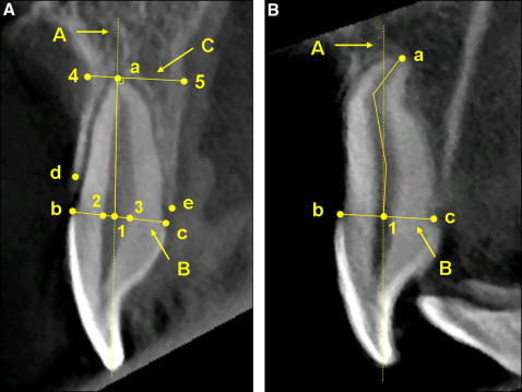
In our study, 60 CBCT records of the 30 patients were collected using a 3-dimensional (3D) x-ray computed tomography device (MCT-1; J. Morita, Osaka, Japan). A voxel size of 0.125 × 0.125 × 0.125 mm was used for all subjects, and depending on the location of the impacted incisor, the other settings were as follows: cylindrical volumes of 4 × 4 cm to 6 × 6 cm, voltage of 80 kV, and current of 5 mA. The slice thickness was set at 1 mm, and the images were reformatted using the i-Dixel software package (version 1.27; J. Morita). On each CBCT record, an author (S.Z.) determined the root development stage of both maxillary central incisors. The measurements used in this study were modified from the reports of Kim et al and Zhong et al. Our reference points, lines, and measurement variables are described in Figure 3 and Table I .

| Measurement variable | Definition |
|---|---|
| RL | Root length: distance from a to 1 |
| RCW | Root canal width: distance from 2 to 3 |
| CEJW | CEJ width: distance from b to c |
| LaBL | Alveolar bone loss of labial side: distance from b to d measured parallel to line A |
| LiBL | Alveolar bone loss of lingual side: distance from c to e measured parallel to line A |
| LaCBT | Alveolar bone thickness of labial alveolar crest: thickness of the alveolar bone 1 mm under point d measured perpendicular to line A |
| LiCBT | Alveolar bone thickness of lingual alveolar crest: thickness of the alveolar bone 1 mm under point e measured perpendicular to line A |
| LaABT | Labial alveolar bone thickness at the root apex: distance from a to 4 measured perpendicular to line A |
| LiABT | Lingual alveolar bone thickness at the root apex: distance from a to 5 measured perpendicular to line A |
| %LaBL | Percentage of LaBL to RL |
| %LiBL | Percentage of LiBL to RL |
| %ABT | Percentage of alveolar bone thickness at the root apex to the CEJ width: LaABT + LiABT/CEJW |
The labial and lingual cementoenamel junction (CEJ) and alveolar crest (the most coronal level of the alveolar bone) were defined on the median sagittal section of the crown, and the apical point was defined as the root apex on a 3D image. Root length was measured from the root apex to the intersection of the long axis of the incisor with the line connecting the labial and lingual CEJ. Alveolar bone loss was calculated from the CEJ to the alveolar crest on the 3D image. If the root was dilacerated, root length and alveolar bone loss were measured in a line following the curvature (the image was rotated as needed). Measurement of the root canal width was made between the intersections of the root canal wall and the line connecting the labial and lingual CEJ. Alveolar bone thickness at the alveolar crest was defined as the thickness of the alveolar bone 1 mm under the alveolar crest, measured perpendicular to the long axis of the incisor on the median sagittal section of the crown. Alveolar bone thickness at the root apex was measured from the root apex to the limit of the alveolar cortex, perpendicular to the long axis of the incisor.
Many previous studies have found that the adjacent permanent teeth used for anchorage can be affected by orthodontic traction. To evaluate changes to the alveolar bone surrounding the anchorage incisors, we used CBCT records to assess pretreatment and posttreatment alveolar bone development for 25 contralateral maxillary central incisors that had reached the occlusal plane before treatment.
All measurements were repeated by 2 investigators (X.S., J.Q.) on 2 separate occasions at 1-week intervals; the average of the 2 measurements yielded the final result. During the follow-up examination when the posttreatment CBCT was taken, the crown heights of the incisors were also measured clinically from the midpoint of the incisal edge to the gingival margin on a line parallel to the long axis of the incisors.
Statistical analysis
For the metric variables, the results are expressed as medians, and means and standard deviations; for nominal variables, they are expressed as frequencies and percentages. The groups were compared using a paired t test or a Wilcoxon signed rank test, as indicated. Statistical analyses were carried out using the SPSS software package (version 17.0; SPSS, Chicago, Ill). All P values are 2-tailed, and the threshold for statistical significance was set at <0.05. The interclass correlation coefficient was 0.99.
Results
Table II gives the mean patient ages at the time of surgery, the sex distribution, the location of impaction, the number of patients with root dilacerations, and the mean durations of closed-eruption treatment, retention, and follow-up. The study sample consisted of 30 patients, 20 boys and 10 girls, with a mean age of 8.44 ± 1.20 years (range, 6.5-11.2 years). There was no statistically significant difference between the male and female subjects in any measurement. Fifteen patients had caries, 8 patients had trauma on their deciduous predecessors, and 7 patients lacked any abnormalities in their deciduous incisors. No patient complained of significant discomfort caused by the therapy.
| Characteristic | n | Mean (SD) | Range |
|---|---|---|---|
| Sex | |||
| Male | 20 | ||
| Female | 10 | ||
| Age (y) | 8.44 (1.20) | 6.5-11.2 | |
| Location of impaction | |||
| Right | 16 | ||
| Left | 14 | ||
| Root dilacerations | 24 | ||
| Mean closed-eruption treatment time (mo) | 10.16 (2.73) | 6.1-14.9 | |
| Mean retention time (mo) | 14.26 (6.07) | 5.0-29.3 ∗ | |
| Mean follow-up time (mo) | 1.76 (3.41) † | 0-15 | |
| Sum of mean retention and follow-up times (mo) | 16.02 (4.81) | 12-29.3 | |
∗ One patient delayed recall time for 12 months, and 4 patients accepted other orthodontic treatments immediately after the closed-eruption treatment.
All 30 impacted immature central incisors were successfully aligned in the dental arches. In 28 patients their root apices had completed development, and in the other 2 patients the apices were at the same stage as those of the contralateral incisors ( Table III ). Reactions of the impacted incisors to cold stimuli were consistent with those of the contralateral incisors. There was no obvious radiographic sign of root resorption or periapical radiolucency. The alveolar bone crest contours of both the labial and lingual sides of the incisors were U-shaped, and no fenestration or dehiscence was found. The mean duration of the closed-eruption treatment (time between applying the power chain and achieving good alignment of the impacted incisor in the dental arch) was 10.16 ± 2.73 months (range, 6.1-14.9 months).
| Stage | Impacted incisors (n) | Contralateral incisors (n) | ||
|---|---|---|---|---|
| Pretreatment | Posttreatment | Pretreatment | Posttreatment | |
| 1 | 8 | 0 | 1 | 0 |
| 2 | 10 | 0 | 9 | 0 |
| 3 | 4 | 0 | 10 | 0 |
| 4 | 7 | 1 | 3 | 1 |
| 5 | 1 | 1 | 7 | 1 |
| 6 | 0 | 28 | 0 | 28 |
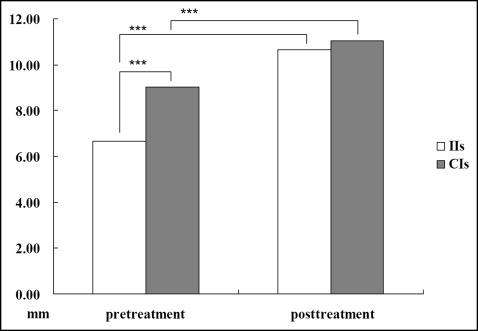
Table IV reports the mean values relating to root and alveolar bone development. Statistically significant differences between the pretreatment and posttreatment root length were found for the impacted incisors ( P <0.001) and the contralateral incisors ( P <0.001). The mean pretreatment root length of the impacted incisors was significantly different from that of the contralateral incisors ( P <0.001), but the difference at posttreatment was not significant ( P = 0.771). The root length ratio of the impacted and contralateral incisors increased after treatment ( P <0.001; Fig 4 ).
| Measurement | Impacted incisors | Contralateral incisors | P | ||||
|---|---|---|---|---|---|---|---|
| Mean (mm) | SD | Range | Mean (mm) | SD | Range | ||
| RL | |||||||
| Pretreatment | 6.67 | 1.94 | 2.47-9.99 | 9.02 | 2.13 | 2.72-12.76 | <0.001 ∗ |
| Posttreatment | 10.66 | 2.10 | 6.98-14.83 | 11.04 | 1.76 | 7.75-13.55 | NS ∗ |
| P | <0.001 ∗ | <0.001 ∗ | |||||
| RCW | |||||||
| Pretreatment | 2.25 | 0.52 | 1.40-3.60 | 2.11 | 0.46 | 1.47-3.32 | NS † , ‡ |
| Posttreatment | 1.97 | 0.35 | 1.33-2.60 | 1.89 | 0.32 | 1.37-2.47 | NS ‡ |
| P | 0.007 † , ‡ | 0.021 † , ‡ | |||||
| LaBL | 2.91 | 1.63 | 0.20-7.05 | 1.40 | 0.91 | 0.00-4.99 | <0.001 † , § |
| LiBL | 0.93 | 1.00 | 0.00-4.00 | 0.77 | 0.68 | 0.00-2.96 | NS † , § |
| P | <0.001 † , § | <0.001 † , § | |||||
| %LaBL | 29.34 | 18.70 | 2.01-64.83 | 13.02 | 8.64 | 0.00-45.49 | <0.001 ∗ , † |
| %LiBL | 9.48 | 12.10 | 0.00-56.26 | 6.97 | 5.97 | 0.00-22.67 | NS ∗ , † |
| P | <0.001 ∗ , † | <0.001 ∗ , † | |||||
| LaCBT | 0.73 | 0.19 | 0.33-1.12 | 0.73 | 0.19 | 0.38-1.00 | NS † , ‡ |
| LiCBT | 1.40 | 0.64 | 0.63-4.19 | 1.20 | 0.41 | 0.71-2.66 | 0.042 † , ‡ |
| P | <0.001 † , ‡ | <0.001 † , ‡ | |||||
| LaABT | 4.24 | 1.97 | 0.18-7.74 | 4.95 | 1.41 | 1.53-7.67 | NS ∗ |
| LiABT | 7.17 | 2.01 | 1.37-11.10 | 6.94 | 1.32 | 4.56-10.53 | NS ∗ , † |
| P | <0.001 ∗ | <0.001 ∗ , † | |||||
| %ABT | 185.34 | 28.79 | 110.11-260.31 | 192.62 | 26.04 | 141.36-247.62 | NS ∗ , † |
| Crown height | 9.87 | 1.16 | 6.99-11.75 | 9.37 | 1.17 | 6.86-11.66 | 0.045 ‡ |


