Enamel

Chapter 32

Enamel

Publisher Summary

This chapter describes dental enamel as a unique biological system that provides a hard surface for the teeth and enables them to reduce food to particles sufficiently small for effective attack by the digestive enzymes. Provided that it remains free from disease, human enamel can withstand a lifetime of crushing work without becoming fractured or completely worn away. Enamel is capable of performing this function only because of its most abundant constituent, hydroxyapatite, which gives it a hardness intermediate between that of iron and carbon steel. Three main aspects of tooth structure determine how physical stress is distributed within enamel. The gross morphology of the enamel cap defines the shape of both the occlusal surface and the amelodentinal junction. Forces within the enamel are transmitted to the underlying dentine, which is softer and more elastic than enamel, and acts as a firm cushion. The internal structure of enamel at histological and ultrastructural levels has an important bearing on its elastic behavior. This concerns the division of enamel into prismatic structures; the shapes, directions, and mutual interlacing of the prisms; and the arrangement of the crystallites within the prisms. In freshly secreted enamel, crystals first appear near the ameloblasts and, as soon as they do so, the enamel takes on the pattern of its final form, an intricate beautifully arranged two-phase system that may not only last throughout life but may also survive in fossil form for millions of years.

Dental enamel is a unique biological system that provides a hard surface for the teeth, and enables them to reduce food to particles sufficiently small for effective attack by the digestive enzymes. Provided that it remains free from disease, human enamel can withstand a lifetime of crushing work without becoming fractured or completely worn away. Enamel is capable of performing this function only because of its most abundant constituent, hydroxyapatite, which gives it a hardness intermediate between that of iron and carbon steel.

If enamel were hard but brittle, as it would be if it was formed from a single crystal of apatite, it would fracture when brought into contact with the enamel on the surface of the opposing tooth, during mastication. This does not occur because enamel has a very high elasticity for a material of its hardness. This elasticity primarily depends upon its being composed of a large number of crystallites, which are in close contact but are not crystallographically continuous, and are arranged in definite patterns. The elasticity of enamel depends on both its own structure, and that of the whole tooth.

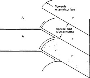
Three main aspects of tooth structure determine how physical stress is distributed within enamel. Firstly, the gross morphology of the enamel cap defines the shape of both the occlusal surface and the amelodentinal junction. Secondly, forces within the enamel are transmitted to the underlying dentine, which is softer and more elastic than enamel and acts as a firm cushion. Thirdly, the internal structure of enamel at histological and ultrastructural levels has an important bearing on its elastic behaviour. This concerns the division of enamel into prismatic structures, the shapes, directions and mutual interlacing of the prisms and finally the arrangement of the crystallites within the prisms (Figure 32.1).

In freshly secreted enamel, crystals first appear near the ameloblasts and, as soon as they do so, the enamel takes on the pattern of its final form, an intricate beautifully arranged two-phase system which may not only last throughout life, but may survive in fossil form for millions of years.

Histological structure

Electron micrographs of mature enamel show that most of its volume is occupied by inorganic crystallites. Though these are discontinuous, they are in such close contact that the second phase, which consists mainly of water with some organic matter, occupies only narrow gaps between the crystals. Early in development the crystals form as long thin ribbons or plates and at this stage a higher percentage of the volume is occupied by the organic phase. The two-phase concept is important (i) in relation to the growth of the inorganic crystallites in the surrounding organic phase during maturation of the enamel and (ii) to the concept of ‘spaces’, which are responsible for the microporous properties of mature enamel.

At a larger order of size, enamel has a prismatic structure, the enamel prism or rod representing the most obvious histologically defined entity. In cross-section its dimensions are approximately the same as those of an ameloblast but it is extremely long and may extend from the amelodentinal junction to the enamel surface. Adjacent prisms are approximately parallel but show local changes of direction. The prisms are formed as the result of the activity of ameloblasts, and one ameloblast may contribute to several prisms, while each prism receives material from more than one cell. In the outermost layer of fully formed human enamel the prisms merge into each other and a thin surface layer is produced. This layer resembles reptile enamel which is less than 1 mm thick and is not prismatic but continuous.

Most of the hydroxyapatite crystals have their long axes approximately parallel to each other and almost parallel to the prism direction (Figure 32.1). Near the edges of the prisms the directions of the crystallites progressively deviate, the ends of the crystallites which are nearer the enamel surface being turned outwards away from the prism axis and towards the prism boundary. As a result of this progressive deviation there is, at the boundary, an abrupt change of crystallite orientation from one prism to the next. The prism boundaries are rendered visible under the microscope by this discontinuity of crystal orientation, rather than as was at one time thought, by a solid prism sheath of organic matter. However, electron micrographs of the earlier stages of enamel maturation do show that the region near the prism boundary contains fewer crystallites and consequently more of the organic phase than within the prisms.

The composition of mature enamel

The physical properties and chemical resistance of enamel are quite different from those of bone, dentine and cementum. At first this appears surprising since all four tissues are mineralized with hydroxyapatite but there are two important differences between enamel and the other tissues. Firstly, whereas bone, dentine and cementum contain some 20% by weight of collagen, mature enamel has only approximately 0·6% of organic matter (Table 32.1) while apatite accounts for approximately 99% of its dry weight. Secondly, the apatite crystals in enamel are approximately ten times wider and thicker (approx. 50 × 50 nm) and much longer than those that impregnate the calcified collagens, so that their volume is at least 1000 times greater. Such large crystals of apatite cannot be produced in synthetic systems at normal temperatures and pressures.

Table 32.1

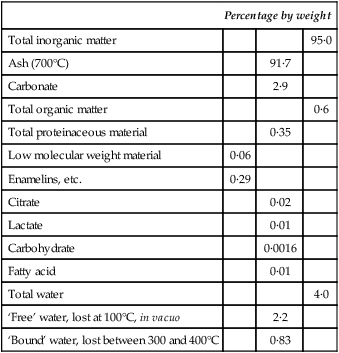
A chemical balance sheet for mature human enamel

  Percentage by weight
Total inorganic matter     95·0
Ash (700°C)   91·7  
Carbonate   2·9  
Total organic matter     0·6
Total proteinaceous material   0·35  
Low molecular weight material 0·06    
Enamelins, etc. 0·29    
Citrate   0·02  
Lactate   0·01  
Carbohydrate   0·0016  
Fatty acid   0·01  
Total water     4·0
‘Free’ water, lost at 100°C, in vacuo   2·2  
‘Bound’ water, lost between 300 and 400°C   0·83  

image

Accurate values for the composition of enamel depend upon the preparation of enamel powder, free from contamination by dentine, surface cuticle, plaque, etc. Contamination is especially serious for investigations of the organic constituents in mature enamel which are present at a very low level. Thus, if enamel with a true content of only 0·5% of organic matter is contaminated by only 1% of its weight of dentine containing 20% of collagen, its apparent content of organic matter would be raised to 0·7%. For this reason the figure of 0·6% for total organic matter in enamel may be slightly high. The value of approximately 0·35% for total ‘true enamel protein’ has been corrected for contaminating collagen on the basis of its hydroxyproline content.

The small amounts of citrate and lactate that are present may be associated with the inorganic phase. The level of carbohydrate in mature enamel is extremely low, the chief sugar components being galactose, glucose and mannose. The fatty acids consist chiefly of palmitate, stearate and oleate in approximately equal amounts.

The water content of enamel is uncertain because of the difficulty of determining the most firmly bound fraction, which is lost only at high temperatures, at which other constituents begin to be decomposed. A total value of 4% by weight for water in mature enamel in vivo is probably realistic. Nuclear magnetic resonance studies have provided evidence for the existence of ‘freely tumbling’ or loosely bound water within the ‘spaces’ of enamel.

Local variations in composition in different regions of the same tooth have been investigated by systematic micro-dissection of 100 μm thick sections of enamel and microanalysis of pieces from specific regions. Another approach is to dissolve the enamel systematically, layer by layer, from the exposed surface and to analyse the resulting solutions. The composition of enamel varies considerably particularly through its thickness. Thus the density of human enamel falls from approximately 3·01 at the surface, where mineralization is highest, to 2·89 near the amelodentinal junction, although local irregularities occur. The inorganic content varies directly, and the organic content inversely, with the density. Carbonate content is also inversely related to density rising from approximately 2·25% at the enamel surface to 3·9% near the amelodentinal junction. Apatite crystals of high carbonate content are less stable than those of low carbonate content as the pH is reduced. This local variation in carbonate content may have some bearing on the pattern of carious attack. Another important local factor affecting caries susceptibility is the distribution of fluoride. This is present at high concentration in the original enamel surface, where it may reach several hundred parts per million, and falls rapidly with depth to 20–50 parts per million in the interior enamel.

The values given in Table 32.1 are expressed on a weight basis whereas in terms of volume about 87·1% is occupied by the inorganic phase, 11·5% by water and 1·4% by organic matter. Mature enamel thus resembles a sintered mass of crystallites with the small spaces between them occupied by water. The small proportion of organic matter is mainly associated with the water in these spaces rather than with the crystallites. Although the aqueous phase occupies a relatively small part of the total volume, it is nevertheless, the continuous phase and is probably in communication throughout the enamel. All the same, in terms of hardness and behaviour under compressive loads, enamel behaves almost as though the inorganic phase were continuous, at least in contrast to dentine and bone where the crystallites are definitely separated by collagen, resulting in these tissues being softer and more deformable under load than enamel.

Gross composition of developing enamel and its changes during maturation

An understanding of the nature of mature enamel and of the protein matrix of enamel can best be obtained by considering (1) the composition of enamel shortly after it has been laid down by ameloblasts and (2) the maturation process which subsequently changes it.

When a tooth first begins to form the enamel appears as a pink jelly-like layer but soon changes to a chalky white mass which, though it looks highly mineralized, is actually quite soft and can be easily cut with a knife or gently scraped away from the more coherent underlying dentine. The enamel remains friable for some time but continuous increase in its degree of mineralization results in its becoming extremely hard before the tooth erupts.

During enamel development several processes occur. Firstly, there is secretion of the organic phase by the ameloblasts, followed by its mineralization; this involves two processes, crystallite formation, occurring soon after secretion of the matrix, and crystal growth, which occurs in several stages. The overall change by which the newly secreted organic phase becomes mineralized is known as maturation.

The changes in composition that developing enamel undergoes during maturation were first studied by Deakins in 1942. He dissected pieces of enamel from pigs’ teeth at various stages of mineralization and measured their contents of inorganic matter, water and organic matter in relation to the increasing density of the enamel, as maturation proceeded.

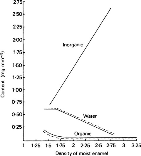
The composition of enamel at the beginning and end of maturation is given in Table 32.2, while the values for inorganic matter, water and organic matter in various intermediate stages of maturation are plotted against density in Figure 32.2.

Table 32.2

Gross composition of pig enamel at the lowest and highest degrees of mineralization

  Lowest mineralization (Very soft; density 1·45) Highest mineralization (Very hard; density 2·76)
  mg mm−3 %by weight Volume* mg mm3 %by weight Volume*
Inorganic 0·54 37·0 0·16 2·62 95·0 0·82
Organic 0·27 19·0 0·20 0·05 1·8 0·04
Water 0·64 44·0 0·64 0·12 4·3 0·12
Total 1·45 100·0 1·00 2·79 101·1 0·98

image

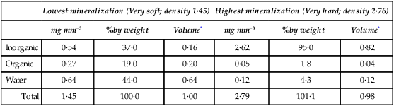
*Volume in mm3 occupied by each constituent, calculated assuming densities of 3·18 for inorganic and 1·31 for organic material

Clearly there is a continuous gain in the inorganic phase and a reciprocal loss of water and organic matter during maturation. Furthermore, as judged by the weight lost per unit volume of enamel, these losses are not simply the result of dilution by the newly forming inorganic crystals but represent the actual removal of both water and organic matter from the tissue. The removal of organic matter during maturation is also apparent from and supported by experiments in which the whole of the enamel from complete deciduous dentitions of human fetuses and newly born infants was separated. It was shown that the total weight of protein present rose to a maximum of approximately 100 mg, 1 month after birth, and then decreased to a steady value which was only one-tenth of this.

The bulk protein of human fetal enamel was subsequently characterized and found to account for 20% of the weight, a value almost equal to that obtained by Deakins for the total organic matter of the youngest pig enamel. Thus the organic matter of young enamel is mainly proteinaceous, only small quantities of other organic constituents, viz. 0·23% of carbohydrate and 0·06% of phospholipid, having been detected.

The total protein of developing enamel has an unusual amino acid composition (Figure 32.3) which distinguishes it from collagens, keratins and the protein of mature enamel. Possession of a unique protein system during its formative stages and the subsequent differential loss of its major protein components during maturation are characteristic features of enamel development which distinguish it from other tissues. The term amelogenins is used to designate those proteins that predominate in newly secreted enamel and which are removed during maturation of the enamel, a process which accompanies its transition from a material with the softness of jelly to one with the hardness of rock. The minor protein components of newly secreted enamel, known as enamelins, are not removed during maturation and largely persist in the fully mature enamel (Figure 32.4). Amelogenins and enamelins contrast strikingly in amino acid composition (cf. Tables 32.4 and 32.5).

Table 32.4

Amino acid composition of amelogenins from various species (values given as residues per 1000 total residues)

Only gold members can continue reading. Log In or Register to continue

  Human Pig Ox
Aspartic acid

Stay updated, free dental videos. Join our Telegram channel

Dec 10, 2015 | Posted by in General Dentistry | Comments Off on Enamel

VIDEdental - Online dental courses

Get VIDEdental app for watching clinical videos