Abstract
The aim of this study was to evaluate the effect of icariin on bone formation during mandibular distraction. 40 Rabbits were randomly divided into experimental and control groups. Mandibular distraction was performed 5 days after unilateral mandibular osteotomy using a custom-made external distractor at a rate of 0.5 mm/12 h for 10 days. From the first day of distraction, icariin (2.5 mg/kg·day) was orally administered to the experimental group and placebo to the controls. 10 Rabbits were killed at the end of weeks 2 and 4 of the consolidation phase. The distracted hemimandible was harvested and newly formed bone was evaluated by soft radiography, histology and bone histomorphometry. Regenerated bone was evaluated for bone mineral density by dual-energy X-ray absorptiometry. The experimental group had fewer radiolucent areas on soft radiography. Bone mineral density of regenerated bone was higher in the experimental than in the control group at 2 and 4 weeks. At 4 weeks, the experimental group had greater volumes of new bone, higher trabecular number, and less trabecular separation than the controls. Oral administration of icariin could promote bone formation during mandibular distraction osteogenesis and might be a promising method for shortening the course of distraction osteogenesis.
Distraction osteogenesis is a common surgical technique that can successfully restore bone defects or lengthen short or hypoplasia bone without the need for bone grafts. Since its first application in the field of oral and maxillofacial surgery by M cCarthy in 1992 , the technique has been used for a variety of craniofacial deformities, including micrognathia, temporomandibular joint ankylosis, obstructive sleep apnea syndrome and bone defects after tumorectomy . The technique is effective in generating new bone by gradually applying tensile strain to an osteotomy site using a distractor. The distraction gap fills with immature bone that differentiates into normal bone after a consolidation phase . Distraction osteogenesis has many advantages over traditional orthopedic techniques, including not requiring bone grafting and simultaneous expansion of the surrounding soft tissue .
Despite the advantages and the general clinical acceptance of distraction osteogenesis, a long consolidation period is required for complete ossification in the distraction gap. Patients are at risk of complications during this time, such as pain, refracture, infection or nonunion , and suffer a greater psychological and economical burden. Many studies have focused on the promotion of new bone formation to shorten the course of distraction osteogenesis. Physical means have included pulsed electromagnetic fields, low-intensity ultrasound and electrical stimulation . Interventional methods, such as transplantation of osteoblast-like cells or bone marrow to the distraction site, injection of growth factors or platelet rich plasma and gene therapy have also been applied to accelerate the maturation of the regenerated bone . Some noninterventional methods, such as administration of calcitonin, alendronate and zoledronic acid have also shown promising results .
Epimedium brevicornum maxim ( Yin Yang Huo , horny goat weed) has been used as a medicinal herb to treat fractures, bone and joint diseases, and gonad dysfunction in traditional Chinese medicine for thousands of years . Icariin (C 33 H 40 O 15 , molecular weight: 676.67), is a major and indicative ingredient isolated from Epimedium brevicornum maxim that has a wide range of pharmacological and biological effects . Its structure shown in Fig. 1 . Icariin-containing drugs exert beneficial effects in the treatment of osteoporosis and prevention of osteonecrosis, through stimulation of bone formation and inhibition of bone resorption . Application of a single component of icariin to a rat hypoandrogenism model showed that icariin might promote bone formation and reduce bone resorption. Icariin has therapeutic potential in the management of hypoandrogenism .The application of icariin during distraction osteogenesis to promote new bone formation has yet not been reported.

In this study, the authors hypothesize that icariin might be a suitable candidate for a non-interventional method to promote bone formation during distraction osteogenesis. The aim of this study is to determine the effect of icariin on bone formation during mandibular distraction by soft radiography, histology, bone histomorphometry and dual-energy X-ray absorptiometry (DEXA).
Materials and methods
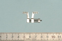
40 Male Japanese white rabbits, 4–5 months old and weighing 3.0–3.5 kg at the beginning of the experiment, were purchased from the Peking University Department of Laboratory Animal Science (Beijing, China). The housing, care, and experimental protocols were approved by the University’s Laboratory Animal Care and Use Committee. The titanium distractors used in this experiment were custom-made and could be fixed to the lateral side of the mandible with three self-tapping titanium screws on each side ( Fig. 2 ). Icariin of 98% purity was commercially purchased from the Tcm Institute of Chinese Materia Medica, NanJing, China.
Surgical procedures
Identical surgical techniques, performed by the same surgeon, were used on every animal. Surgery was performed under general anesthesia with an intravenous injection of 30 mg/kg pentobarbital sodium (Sigma, USA) and subcutaneous administration of 2% lidocaine at the surgical site. The submandibular and cheek hairs were shaved. A submandibular curve incision was made in each rabbit along the inferior border of the right mandible and the periosteal flap was raised from the inferior surface of the mandible body. Unilateral osteotomy was performed with a water-cooled fissure burr just between the first premolar and the mental foramen. The osteotomy was completed using a small osteotome. Custom-made external titanium distractors were applied and secured to the mandible with self-tapping screws. The distractor was activated during the operation to confirm the successful osteotomy, then rotated back to the original position. Subcutaneous tissues and skin were closed in layers ( Fig. 3 ).

The animals were housed in separate cages in a recognized animal holding facility on a 12-h light/dark schedule and were fed ad libitum . 40,000 IU/kg penicillin and 80,000 IU/kg streptomycin were administered intramuscularly daily for 5 days. The animals’ body weights were monitored every week postoperatively to adjust the administration of icariin.
Mandibular distraction and icariin administration
All rabbits were randomly divided into two groups (experimental and control), with 20 rabbits in each. Following a 5-day latency period, the distraction was performed at a rate of 0.5 mm twice daily for 10 days, to a total length of 10 mm. From the first day of the distraction, icariin (2.5 mg/kg·day) was orally administered to the experimental group and a placebo to the control group until the animals were killed. At the end of the second and the fourth week of the consolidation phase, 10 animals from each group were killed with an intravenous overdose of 100 mg/kg pentobarbital. The distracted hemimandible was harvested and the surrounding soft tissue was excised.
The distances achieved were measured using a sliding caliper (mean of middle, superior and inferior boundaries). The newly formed bone in the distracted gap was evaluated by soft radiography, histology, bone histomorphometry and DEXA.
Radiographic evaluation and DEXA
The mandibles were cut into left and right halves. Lateral soft X-ray (REGIUS PureView System, Konica Minolta, Japan) radiographs were taken. The bone mineral density (BMD) of the newly formed bone was determined using DEXA (Lunar Prodigy, General Electric, USA). The distracted mandible was scanned and analyzed using the special animal program provided by the manufacturer. All experimental data were sampled three times.
Histology and histomorphometric analysis
The distracted bones and the adjacent original bone were cut out en bloc and split longitudinally along the median axial plane. The specimens were fixed in phosphate-buffered 10% formal saline for 24 h, decalcified with 14% EDTA for 5–6 weeks, then dehydrated in an ethanol series and embedded in paraffin. Sections of 5 μm were cut, one section was selected per 50 μm, five selected sections of each specimen were included and stained with hematoxylin–eosin. Digital images of histological sections were captured using a light microscope (OP750, OLYMPUS, Japan) and histomorphometric measurements were made using a computerized image analysis system (Leica QWin Plus, Leica, Germany) for blind quantification of new bone formation in the distracted gap. The nomenclature and calculations were in accordance with the American Society of Bone and Mineral Research Histomorphometry Nomenclature Committee .
Statistical analysis
Mean values and standard errors of the mean are presented. A one-way analysis of variance (ANOVA) was used to calculate differences of the distracted length and an unpaired t test (two tailed) was used to compare differences in BMD and histomorphometric parameters of each group. All statistical analyses were carried out using SPSS software 11.0 (SPSS, Munich, Germany). The level of statistical significance was set at P < 0.05.
Results
Clinical evaluation
Both the surgical procedure and the distraction period were well-tolerated by all rabbits. The lengthening of the mandible caused malocclusion and overgrowth of upper and lower incisors. In order not to affect mastication and protect the gingiva, the incisors were shortened by regular grinding. Gross postdistraction specimens clearly demonstrated evidence of callus formation at the site of distraction in both groups. The average distraction length for all rabbits was 9.66 ± 0.30 mm.There were no statistical differences in the mean distraction length between the experimental and the control groups (ANOVA, F = 0.890; df = 39; P > 0.05).
Examination of soft X-ray radiographs
Representative soft X-ray radiographs of each group are shown in Fig. 4 . At 2 weeks, the soft X-ray radiographs of both groups showed radiolucent and slightly radio-opaque areas in the distracted gaps. The radiodensity in the experimental group appeared to be higher than in the control group, although it was not significant. At 4 weeks, radio-opacity was observed in most of the distracted gaps in both groups and was more pronounced in the experimental group than that in the control group ( Fig. 4 ).
Bone densitometry
The BMD of the regenerated bone is shown in Fig. 5 . Analysis of the bone in the distraction gap revealed that BMD in the experimental group was higher than in the control group, at 2 weeks (0.237 ± 0.040 g/cm 2 vs. 0.190 ± 0.057 g/cm 2 , t test, t = 2.130; df = 18; P < 0.05) and at 4 weeks (0.295 ± 0.045 g/cm 2 vs. 0.245 ± 0.058 g/cm 2 , t test, t = 2.185; df = 18; P < 0.05). With the administration of icariin, the BMD of regenerated bone in the distraction gap significantly increased, by 25% and 20% after 2 and 4 weeks of consolidation, respectively.
Histology and histomorphometric analysis
After 2 weeks of consolidation, sparse trabeculae surrounded by proliferating osteoblasts arranged along the direction of distraction could be seen in the distracted gaps of both groups. Osteoid could be observed on the surface of newly formed trabecular bone. Bone fusion could clearly be seen between the regenerated bone and the adjacent original bone. At the end of 4 weeks of consolidation, the trabecular zone increased and became thicker, and the boundary between the regenerate bone and the adjacent original bone and the adjacent original bone became unclear. Newly formed trabeculae, aligned parallel to the axis of lengthening, became joined by bridges ( Fig. 6 ). More cartilaginous islands could be observed in the control group than in the experimental group at both time points.


