Diagnostic Case I:
Tooth Fracture: Unrestorable
Suanhow Howard Foo

Chief Complaint
“I had excruciating pain last night, now I can’t touch my tooth.”
Medical History
The patient (Pt) was a 58-year-old male Caucasian. He presented with nothing significant in medical history and no allergies to any medications or to latex. Vital signs were: Blood pressure (BP) 132/87 mmHg, pulse 82 beats per minute (BPM), respiratory rate (RR) 17 breaths per minute.
The Pt was American Society of Anesthesiologists Physical Status Scale (ASA) Class II.
Dental History
Pt had on-and-off pain on the lower right quadrant for a few weeks and was referred for an evaluation of tooth #31. The tooth had a mesial (M) to distal (D) crack. The tooth was painful to touch and the Pt could not eat or bite on that tooth. Pt reported a history of bruxism.
Clinical Evaluation (Diagnostic Procedures)
Examinations
Extra-oral Examination (EOE)
No asymmetry, no lymphadenopathy, no deviation of jaw when opening, no swelling, and temporomandibular joint (TMJ) was within normal limits (WNL).
Intra-oral examination (IOE)
Oral cancer screening performed with all tissues WNL. Tooth #31 had a M to D crack. Periodontal exam showed probing depths from M to D of Facial (4 mm, 3 mm and 8 mm) and M to D of Lingual (4 mm, 4 mm and 8 mm). Tooth #31 had type 1 mobility. Tooth #30 had probing depths from M to D of Facial (4 mm, 3 mm and 4 mm) and M to D of Lingual (4 mm, 4 mm and 4 mm). Tooth #31 had pain with bite test and pain when occluding. Methylene blue dye and fiber optics showed fracture was through and through and extended below the cementoenamel junction (CEJ).
Diagnostic Tests
| Tooth | #29 | #30 | #31 |
| Percussion | – | – | + |
| Palpation | – | – | – |
| Cold | Normal | Normal | – |
| Mobility | None | None | Class 1 |
| Bite | – | – | + |
+: Response to percussion, or bite stick test;
– : No response to percussion, palpation, cold, or on bite stick test
Radiographic Findings
Tooth #31 had a radiolucency that extended from the D cervical area to the apex of the D root. A crack could be seen on the D portion of tooth #31 with the D restorative material fractured. (See Figures 2.1 and 2.2.)
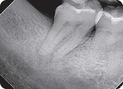
Figure 2.1 The initial radiograph of tooth #31. Notice the shallow restoration and the periapical rarefaction at the root apices.

Figure 2.2 The extent of rarefaction in the distal root of tooth #31. Note how the radiolucency moves up to the alveolar crest.
Pretreatment Diagnosis
Pulpal
Pulp Necrosis, tooth #31
Apical
Symptomatic Apical Periodontitis, tooth #31
Treatment Plan
Recommended
Emergency:Extraction, tooth #31
Definitive:Extraction, tooth #31
Alternative
No treatment
Restorative
Implant or Fixed Prosthetics
Prognosis
| Favorable | Questionable | Unfavorable |
| X |
Clinical Procedures: Treatment Record
First visit (Day 1): Exam: Pt was referred for an evaluation of tooth #31. Medical history (Hx) and vital signs were taken. Three periapical (PA) radiographs were prescribed in order to evaluate the PA area for possible infection and to determine the extent of the crack. The radiographs showed PA rarefactions (Figures 2.1 and 2.2) at root tips and bone loss in D root area. Clinical tests and exams were performed. Tooth #31 had an M to D crack that was verified with methylene blue (Figure 2.3) and a fiber optic light (Figures 2.4 and 2.5). The tooth could be separated in a buccal–lingual (B–L) manner with light touch. The defect could be seen extending to the pulpal floor. Pt was informed that the prognosis of the tooth was unfavorable and that extraction was needed to alleviate his pain and for healing to occur. The Pt accepted treatment (Tx) of extraction of Tooth #31. The extracted tooth was photographed and confirmed the initial diagnosis of a root fracture and split tooth (Figure 2.6).

Figure 2.3 Mesial to distal crack of tooth #31, stained with methylene blue to better visualize the extent of the crack.

Figure 2.4 Fiber optic light illumination of tooth #31 shows that the crack goes below the CEJ. The light does not pass through from lingual to buccal.

Figure 2.5 Fiber optic light was used on the buccal surface to confirm the crack.

Figure 2.6 Diagnosis of a split tooth is confirmed after the extraction of tooth #31.
Post-Treatment Evaluation
Second visit (1-week follow-up): Pt returned for a post-operative (PO) follow-up. The area around the extraction site of tooth #31 was neither inflamed nor swollen. Gingival tissue had already begun to fill in the socket. The Pt was able to eat and brush his teeth in the lower right quadrant.


