Introduction
The aims of this study were to investigate the relationship between dental age and maxillary canine displacement with 2 methods, to study differences in dental development between the displaced and nondisplaced sides, and to investigate sex differences in dental development in the studied groups.
Methods
Two hundred forty subjects were recruited for this study. They were divided into 2 groups: the palatally displaced canine group (87 girls, 33 boys) and the buccally displaced canine group (81 girls, 39 boys). Dental ages were assessed by using the 2 methods.
Results
Delayed dental development was associated with subjects with palatally displaced canines only, whereas those with buccally displaced canines had comparable dental development to that of the control group. Sex differences in the 3 groups were found; girls had more pronounced delayed dental development in all groups according to both methods. With unilateral palatally displaced canines, the displacement side showed delayed dental development compared with the side of nondisplacement ( P = 0.002). The development rates of the displaced and nondisplaced sides were comparable in the subjects with unilateral buccally displaced canines.
Conclusions
Palatal and buccal canine displacements are 2 distinct entities; delayed dental development plays a role in the etiology of palatal canine displacement, but not in buccal canine displacement.
The ectopic eruption of the maxillary permanent canine, whether it results in subsequent impaction or displacement, is a frequently encountered clinical problem. Different theories have been proposed to explain the etiology of maxillary canine impactions, but the exact etiology remains obscure. Palatally and buccally impacted or displaced canines are considered 2 completely different phenomena, with the etiology for one differing from the other.
Late-developing dentitions with insufficiently developed lateral incisors can affect the migration of the developing canine and result in its palatal impaction. Becker and Chaushu examined the dental ages of 47 patients with buccally and 55 with palatally ectopic canines. They assessed dental age from panoramic and periapical radiographs of the incisor region and reported that half the subjects with palatal displacements showed retarded dental development, and those with buccal displacements were not associated with any unusual retardation and were comparable with the control group. More recentlty, Różyło-Kalinowska et al linked delayed dental development to impacted canines. They reported that dental development in patients with impacted canines, either buccally or palatally, was delayed when compared with the control group. However, they found no differences in dental development between buccally and palatally impacted canines.
Many methods have been introduced to determine dental age. Dental age can be determined by either the stage of emergence or the stage of tooth formation. Tooth formation has many advantages over emergence when used to evaluate maturity and estimate a patient’s age. These include the ability to study the majority of teeth in 1 radiographic examination with the tooth-formation method, whereas tooth emergence is only a short phase of the process of tooth eruption, which limits the number of teeth available to study. With the tooth-emergence method, only the teeth visible in the oral cavity can be considered and studied, and these might be limited to just a few teeth. Emergence can only be used up to the age of 30 months, after the deciduous teeth are completed, and after 6 years, after the eruption of the first molar, which limits its applicability. Another drawback of using the eruption of teeth is that the timing of emergence differs from 1 child to another. In addition, several local, external, and environmental factors influence the emergence of teeth—eg, early loss of the predecessor, space conditions in the arch, ankylosis, impaction, and some syndromes. However, an advantage of tooth eruption is its simplicity and ease of use. Based on these facts, using the formation and calcification of teeth to determine dental age is a much more accurate, precise, and reliable indicator of dental maturity than tooth emergence.
Delayed dental development has been linked to palatally displaced canines, but the research in this area until now has been minimal. The aims of this study were to (1) investigate the relationship between dental age and maxillary canine displacement by using the methods of Demirjian et al and Nolla, (2) investigate sex differences in dental development in the studied groups, and (3) study differences in dental development between the displaced and nondisplaced sides.
Material and methods
The records of 3000 patients available in the archives of the Dental Teaching Center of Jordan University of Science and Technology and other private orthodontic practices were screened by an investigator (D.H.N.) for buccal or palatal displacement of the maxillary canine. Those with diagnosed ectopic canines were selected. The type of displacement was diagnosed by using the subjects’ dental records (intraoral radiographs, file notes, and study casts). Dental records for all subjects were available as part of their comprehensive orthodontic treatment.
A total of 240 orthopantomograms with displaced canines were found for this study. Although the diagnosis of displaced canines cannot be determined earlier than 10 years, some subjects who were diagnosed with canine displacement later (after 17 years) had orthopantomograms in their files taken at early stages for other reasons. These orthopantomograms were included in the study.
The subjects were divided into 2 groups.
Group 1 included those with palatally displaced canines. The canine was defined as palatally displaced if it was positioned palatally to the line of the arch, whether it was impacted or erupted. This group consisted of 120 subjects (87 girls, 33 boys). Their ages ranged from 8 to 17 years (average, 13.92 ± 2.00 years). The subjects in this group had unilateral (83 subjects) or bilateral (37 subjects) palatally displaced canines.
Group 2 included subjects with buccally displaced canines. The canine was defined as buccally displaced if it was positioned buccally to the line of the arch, whether it was impacted or erupted. This group consisted of 120 subjects (81 girls, 39 boys). Their ages ranged from 7 to 16 years (average, 12.79 ± 1.9 years). The subjects in this group had unilateral (58 subjects) or bilateral (62 subjects) buccally displaced canines.
The subjects selected for this study fulfilled the following criteria: (1) white origin, (2) chronologic age up to 17 years when the orthopantomogram was taken, (3) clear panoramic radiographs, (4) no aplasia of 2 corresponding teeth bilaterally in the maxilla or the mandible, and (5) no syndromes or relevant serious medical abnormalities.
One hundred twenty subjects (80 girls, 40 boys) with no maxillary canine displacements were selected as the control sample. Their ages ranged from 8 to 16 years (average, 13.34 ± 1.94 years).
Chronologic age for each subject was obtained by subtracting the birth date from the date of the orthopantomogram. The chronologic age in months was converted into a decimal age by dividing the number of months by 12.
Dental age was assessed by using the methods of Demirjian et al and Nolla. With the method of Demirjian et al, the 7 left mandibular teeth (excluding the third molars) were assessed in each orthopantomogram. When a tooth was missing on the left side, the corresponding tooth on the right side was used for the assessment. Each tooth was given a stage from A to H by following the criteria described in writing and by comparing the tooth to the diagrams in the original article. Stages assigned for each of the 7 left mandibular teeth were converted into maturity scores with the conversion table. The scores were then added together, and the total score for each subject was converted into dental age by using the 2 tables of standards for boys and girls by Demirjian et al.
With the method of Nolla, both maxillary and mandibular teeth were assessed from the orthopantomogram. The development of a particular tooth was appraised by matching it as closely as possible to the illustration provided by Nolla, and a stage from 0 to 10 was given for each tooth. When the radiographic reading of the tooth’s development lay halfway between 2 stages, the reading was given a value of 0.5. When the radiographic development showed a reading that was slightly greater than the illustrated grade, but not as much as halfway between that stage and the next, the reading was given a value of 0.2. When the radiograph showed a stage of development that was near the next stage but had not quite reached it, a value of 0.7 was added to the reading. The obtained scores for the maxillary and mandibular teeth on each side, right and left, were added separately. Third-molar scores were included only in patients older than 16 years of age. The sum of scores of the maxillary and mandibular teeth on each side were converted into dental ages by using Nolla’s tables of norms for boys and girls for the 14 mandibular and maxillary teeth combined if the third molars were not included, and the tables for the 16 maxillary and mandibular teeth if the third molars were included. The dental ages for the right and left sides of the same subject were recorded separately. The measurements were made by the same investigator (D.H.N.) in a period of 1 month.
Twenty orthopantomograms were randomly selected, and the dental ages with the methods of Demirjian et al and Nolla were rescored after an interval of 2 weeks to test intraexaminer reliability with the kappa test. The kappa values were above 92%, which indicated substantial agreement between readings.
Statistical analysis
The data were analyzed with the Statistical Package for the Social Sciences computer software (version 17.0; SPSS, Chicago, Ill).
Descriptive analyses (means, standard deviations) for chronologic and estimated dental ages by using the methods of Demirjian et al and Nolla (left-side measurements only) in the 3 groups were calculated. The Student t test was used to detect differences between the chronologic and the estimated dental ages in the 3 groups. In unilateral displacements, dental ages assessed by Nolla’s method between the displaced and nondisplaced sides were compared with paired t tests. Significance was predetermined at the 0.05 level.
Results
The means, standard deviations, and differences between the means and P values of the chronologic and estimated dental ages by using the methods of Demirjian et al and Nolla in girls, boys, and total sample of the 3 groups are presented in Tables I and II .
| Group | Girls | Boys | Total | ||||||
|---|---|---|---|---|---|---|---|---|---|
| Chronologic age | Dental age (Demerjian et al ) | Dental age (Nolla ) | Chronologic age | Dental age (Demerjian et al ) | Dental age (Nolla ) | Chronologic age | Dental age (Demerjian et al ) | Dental age (Nolla ) | |
| PDC | 13.92 ± 2.01 | 13.30 ± 1.56 | 12.83 ± 1.80 | 13.92 ± 2.03 | 13.44 ± 1.95 | 13.20 ± 2.00 | 13.92 ± 2.00 | 13.34 ± 1.67 | 12.93 ± 1.86 |
| BDC | 12.67 ± 1.92 | 12.70 ± 1.63 | 12.13 ± 1.80 | 13.03 ± 1.84 | 13.13 ± 1.33 | 13.02 ± 1.30 | 12.79 ± 1.90 | 12.84 ± 1.55 | 12.42 ± 1.70 |
| Control | 13.32 ± 1.96 | 13.25 ± 1.65 | 12.83 ± 1.73 | 13.39 ± 1.91 | 13.45 ± 1.62 | 13.28 ± 1.71 | 13.33 ± 1.94 | 13.31 ± 1.64 | 13.00 ± 1.73 |
| Total | 13.32 ± 2.02 | 13.08 ± 1.63 | 12.60 ± 1.80 | 13.42 ± 1.94 | 13.34 ± 1.63 | 13.16 ± 1.67 | 13.35 ± 2.00 | 13.16 ± 1.63 | 12.78 ± 1.78 |
| Age | PDC | BDC | Control | ||||||
|---|---|---|---|---|---|---|---|---|---|
| Correlation | Mean difference (SE) | P value | Correlation | Mean difference (SE) | P value | Correlation | Mean difference (SE) | P value | |
| Girls | |||||||||
| Chronologic-Demerjian et al | 0.87 ∗ | 0.61 (0.11) | 0.000 | 0.86 ∗ | −0.02 (0.11) | 0.831 | 0.89 ∗ | 0.07 (0.10) | 0.484 |
| Chronologic-Nolla | 0.89 ∗ | 1.09 (0.10) | 0.000 | 0.89 ∗ | 0.54 (0.10) | 0.000 | 0.88 ∗ | 0.48 (0.11) | 0.000 |
| Boys | |||||||||
| Chronologic-Demerjian et al | 0.89 ∗ | 0.48 (0.16) | 0.005 | 0.78 ∗ | −0.11 (0.19) | 0.570 | 0.91 ∗ | −0.06 (0.13) | 0.618 |
| Chronologic-Nolla | 0.90 ∗ | 0.73 (0.16) | 0.000 | 0.77 ∗ | 0.01 (0.19) | 0.968 | 0.91 ∗ | 0.11 ± 0.12 | 0.387 |
| Total sample | |||||||||
| Chronologic-Demerjian et al | 0.87 ∗ | 0.58 (0.09) | 0.000 | 0.84 ∗ | −0.05 (0.09) | 0.594 | 0.90 ∗ | 0.03 (0.08) | 0.742 |
| Chronologic-Nolla | 0.89 ∗ | 0.99 (0.08) | 0.000 | 0.84 ∗ | 0.37 (0.09) | 0.000 | 0.88 ∗ | 0.36 (0.08) | 0.000 |
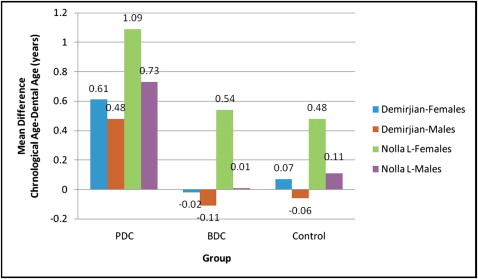
The mean differences between the chronologic and dental ages estimated by the methods of Demirjian et al and Nolla in the 3 groups are compared in Figure 1 . The comparison showed that the delay in dental age was more pronounced in the palatally displaced canine group; dental ages were delayed according to both methods, and the statistical significance of this delay was higher in this group compared with the buccally displaced canine and the control groups.
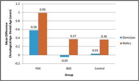
Subjects in the palatally displaced canine group exhibited a significant delay in their dental age, and both methods underestimated the true chronologic ages ( P <0.001). The delay in dental age was greater with the method of Nolla ; the mean difference with Nolla (0.99 years) was greater than that with the method of Demirjian et al (0.58 years). The statistical significance of the delay was higher in Nolla’s method ( t = 11.635) than in the method of Demirjian et al ( t = 6.403). The delay in dental development was greater and more significant in girls (0.61 and 1.09 years with the methods of Demirjian et al [ P <0.001] and Nolla [ P <0.001], respectively) than in boys (0.48 and 0.73 years with the methods of Demirjian et al [ P <0.01] and Nolla [ P <0.001], respectively) ( Fig 2 ).