Figure 2.25 (a) Panoramic radiography illustrates dental and periodontal status and indicates pathology of right coronoid process. (b) CT showing hyperplasia of the right coronoid process interfering with the zygomatic arch.
I. Diagnosis/Diagnoses
Expanded DC/TMD
- Coronoid hyperplasia.
J. Case Assessment
- There were nutritional challenges because of problems with opening mouth and chewing. The patient had chosen to eat food of low nutritional value during the period of restricted jaw opening.
- After a long time with restricted mandibular opening the masticatory muscles may have atrophied. Therefore, comprehensive physical therapy may be needed, both by physical therapist and self-assisted training.
- Coronoid hyperplasia may reappear after surgical excision.
K. Evidence-based Treatment Plan including Aims
Treatment goals
- To achieve the possibility to open jaw normally without pain.
- Dental hygiene instructions.
- Physical therapy.
Management
- Coronoidotomy of the right coronoid processes.
- An intraoral approach was performed where the right coronoid process was exposed. A coronoidotomy where the hyperplastic coronoid was sectioned and a 5 mm segment was resected.
- Jaw exercises.
L. Prognosis and Discussion
- Surgery is the standard treatment option with the goal to eliminate mechanical obstruction of the coronoid process interfering with the zygomatic arch at mouth opening. An intraoral approach was chosen to minimize scares and risk of facial nerve damage. Either coronoidectomy or coronoidotomy are performed. If coronoidectomy is performed the coronoid process is resected, whereas in a coronoidotomy the process is sectioned and left. Less postoperative morbidity has been reported in cases with a coronoidotomy; on the other hand, there is a higher recurrence rate due to risk of reattachment of the coronoid process.
- No facial nerve paresthesia was observed after the operation. The occlusion was stable with no vertical anterior or horizontal open bite. Postoperative physiotherapy was instituted the first weeks with stretching exercises for the preservation of the increased mouth opening. At 6 months postoperatively the patient had increased maximal mouth opening (38 mm).
- Reattachment of the coronoid process and regeneration of the coronoid process after coronoidectomy or coronoidotomy might occur. In this case, radiological findings 3 years after surgery showed regrowth and a new hyperplasia of the coronoid process (Figure 2.26).
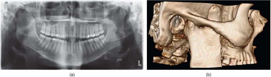
Figure 2.26 (a) Panoramic radiography 3 years after coronoidotomy shows regeneration of the right coronoid process. (b) CT showing regeneration of the coronoid process 3 years after coronoidotomy and a new hyperplasia of the right coronoid process.
Background Information
- Coronoid hyperplasia is a rare condition that may affect one or both coronoids.
- The etiology is unknown, but muscular hyperactivity or trauma may be reasons for the hyperplasia. In cases with condylar destruction one may observe coronoid elongation or hyperplasia.
- The etiology of mandibular hypomobility can be classified into:
- intracapsular – this group may have internal derangement, degenerative arthritis, intracapsular fracture, infection;
- extracapsular, like coronoid hyperplasia – other causes include, muscle contracture, radiation, fibrosis, scarring from trauma or prior surgery;
- neurologic (e.g., traumatic brain injury, tetany);
- psychogenic, hysterical trismus or conversion reaction.
- Decreased mouth opening capacity as one of the clinical signs may appear gradually. Pain may or may not be present.
Stay updated, free dental videos. Join our Telegram channel
VIDEdental - Online dental courses


