Introduction
Fixed retainers are widely used after orthodontic treatment, sometimes for extended periods, despite insufficient knowledge of their possible long-term adverse effects on the periodontium. The aim of this study was to evaluate whether bonded orthodontic retainers have an adverse long-term effect on the marginal bone levels of the mandibular front teeth.
Methods
The study included 62 consecutive patients in 3 groups: (1) patients who underwent orthodontic treatment and wore a fixed retainer for 10 years, (2) patients who underwent orthodontic treatment but did not have a fixed retainer, and (3) untreated controls. The marginal bone levels were measured by cone-beam computed tomography 10 years after treatment. Additionally, multivariate data analysis was used to analyze possible correlations between the marginal bone levels at 10 years and the variables obtained from the study casts and profile radiographs.
Results
The results demonstrated a significantly lower marginal bone level on the buccal side of the mandibular front teeth in the orthodontically treated patients compared with the orthodontically untreated group. There was no difference in the marginal bone levels between the retainer group and the no-retainer group. Multivariate analysis indicated that a low marginal bone level was correlated with a basal open vertical relationship, posterior rotation of the mandible, pretreatment of the incisor protrusion, and extraction therapy.
Conclusions
Within the limits of this research design, the long-term retention phase in general does not seem to cause any adverse effects on the marginal bone levels after 10 years.
Highlights
- •
Long-term bonded lingual retention does not adversely affect marginal bone level.
- •
Orthodontic patients had lower marginal bone levels buccally on the mandibular front teeth.
- •
Low marginal bone level was correlated with a basal open vertical relationship.
- •
It was also correlated with mandibular posterior rotation in orthodontic patients.
Only 10% of orthodontically treated patients maintain clinically acceptable mandibular alignment for up to 20 years postretention. This is due to relapse caused by the remodeling of the periodontal tissues and long-term changes in the occlusion from facial growth, dentoalveolar development, and function. No clear predictors for stable long-term mandibular alignment have been identified, and it has been concluded that orthodontic stability cannot be predicted at the individual level. As a result, many orthodontists routinely provide retention for their patients in the form of bonded lingual retainers at the end of active orthodontic treatment. When regular monitoring is performed, bonded lingual retainers are considered to be an efficient, effective, and reliable option for long-term use.
Although bonded lingual retainers are widely used, current knowledge of their possible adverse effects on oral health is low. With regard to the effect of bonded lingual retainers on the periodontium, previous studies according to a recent systematic review provide contradictory results and a low level of evidence. Several studies have indicated that bonded lingual retainers promote calculus accumulation, gingival inflammation, increased bleeding on probing, gingival recession, and increased pocket depth. Other studies concluded that there is no negative long-term effect of bonded lingual retainers on periodontal health. Except for the above, other factors should be considered for the long-term use of bonded lingual retainers. Teeth normally have physiologic mobility during chewing; furthermore, mesial migration takes place over time. The use of a bonded lingual retainer maintains the teeth as a unit, which is a type of splinting. The question remains whether this splinting, which reduces the mobility of the teeth, is harmful for the periodontium over time. Moreover, is it possible that the splinting, combined with the mesial migration of the teeth in the lateral segments, forces the anterior segment “in block” out of the bone envelope with a possible increase of the risk for buccal recession? It is still unclear whether bonded lingual retainers have any long-term adverse effects on the periodontium, especially on the marginal bone level. So far, only intraoral periapical radiography has been used to evaluate the marginal bone levels around teeth “splinted” with bonded lingual retainers, with the disadvantage of only exploring the mesial and distal tooth surfaces.
The purpose of this study was to assess the potential adverse effects of bonded mandibular lingual retainers on the periodontium of the mandibular front teeth after 10 years by measuring the alveolar bone levels in tomographic images by using cone-beam computed tomography (CBCT) for the assessment. The null hypothesis was that there is no difference in the marginal bone levels between orthodontic patients with long-term bonded lingual retainers compared with patients without retainers and untreated controls.
Material and methods
This study had a cross-sectional design regarding the primary outcome and the marginal bone levels, and a longitudinal design regarding the possible predictive variables ( Table I ). Patients who had finished treatment between 1998 and 2002 at the Clinic of Orthodontics, Public Dental Service in Gothenburg, Sweden, were recalled for a follow-up examination.
| Retainer group (OR) n = 34 |
No retainer group (O) n = 14 |
Control group (C) n = 14 |
|
|---|---|---|---|
| Pretreatment (T0) | Study casts Profile radiograph | Study casts Profile radiograph | |
| Posttreatment (T1) | Study casts Profile radiograph | Study casts Profile radiograph | |
| 5 years posttreatment (T5) | Study casts | Study casts | |
| 10 years posttreatment (T10) | Study casts Profile radiograph CBCT | Study casts Profile radiograph CBCT | Study casts Profile radiograph CBCT |
The inclusion criteria were nonsmoking adolescent patients with no signs of periodontal disease, treated in both arches with fixed appliances (MBT) and preadjusted appliances (both, 3M Unitek, Monrovia, Calif) with 0.022-in slots and with available pretreatment and posttreatment data, study models, and profile radiographs.
Furthermore, patients in the retainer group had mandibular multistrand lingual retainers (PentaOne 0.0195; Masel Ortho Organizers, Inc, Carlsbad, CA) bonded to the 6 front teeth (canine to canine).
The exclusion criteria were adults who had orthodontic treatment in 1 jaw and removable appliances; patients who had surgery, agenesis, or impacted canines; or patients who declined to participate in the study or sign a consent agreement.
Moreover, a third group (control group) of orthodontically untreated periodontally healthy subjects, matched by age, sex, and malocclusion to the orthodontically treated groups at the 10-year follow up, was sampled from people at the University of Gothenburg in Sweden.
Of the 62 patients included in the study, 34 had orthodontic treatment followed by a retention period with a bonded mandibular lingual retainer for 10 years (OR), 14 had orthodontic treatment with short retention times less than 5 years (O), and 14 were control subjects with no orthodontic treatment (C). The mean age of all patients was 27 years at the 10-year follow-up; 66% were female, and 53% were classified as Angle Class I, 45% as Angle Class II, and 2% as Angle Class III. The patients were evenly distributed between the groups regarding age, sex, and malocclusion. Among the orthodontically treated patients, 69% had orthodontic therapy that involved the extraction of teeth in both arches; they were equally distributed between the malocclusion groups, and the rest of the patients had no extractions.
Ethical approval for the study was obtained from the Research Ethical Board at the University of Gothenburg. The Swedish Commission on Radiological Protection approved all radiographs (Dnr 290-09).
The following were the study variables.
- 1.
Outcome variable. The primary outcome variable was the marginal bone level.
- 2.
Predictor variables. The primary predictor variable of interest in this study was the presence of a bonded mandibular lingual retainer, 10 years posttreatment. The secondary predictor variables were incisor irregularity and the inclination (ILi/ML and ILi/NB) and position (Ii-B and Ii-APg) of the mandibular incisors at pretreatment, posttreatment, and 10-years posttreatment. Other predictor variables of interest were extraction or nonextraction therapy, Angle classification, and the sagittal (SNA, SNB, and ANB) and vertical relationships (NSL/ML, NSL/ML, and NL/ML) of the patient.
- 3.
Other variables. Age and sex.
The treatment data, study models, and profile radiographs were collected from the orthodontically treated group before treatment, directly after treatment, and 10 years posttreatment (T0, T1, and T10). CBCT radiographs were collected from the subjects in all groups at T10 ( Table I ).
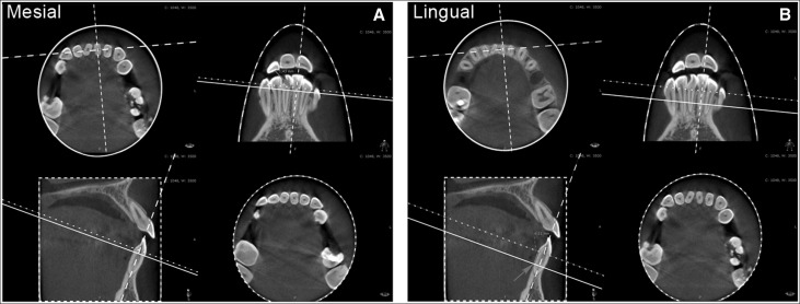
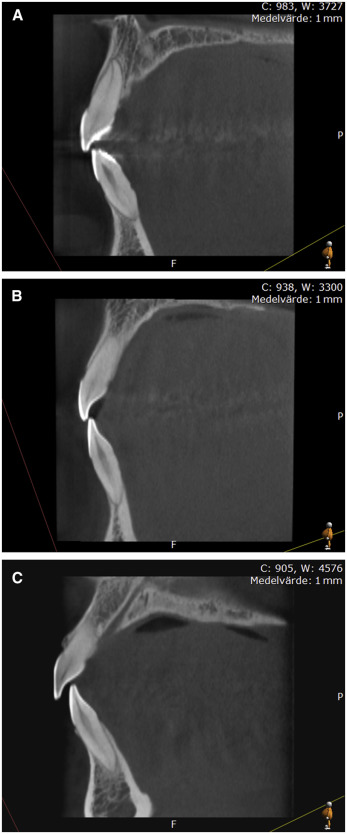
Marginal bone levels were measured as follows. A CBCT examination was performed using a 3DX Accuitomo FPD (J. Morita, Kyoto, Japan) with a 60 × 60-mm field of view, 360° rotation, and exposure parameters of 17.5 seconds, 75 kV, and 4-5, 5 mA (milliampere). The image volume was reformatted using software (i-Dixel-3DX, 3D, version 1.691; J. Morita), the Sectra IDS5 (Sectra Imtec AB, Linköping, Sweden), and the PACS Multi Planar Reconstruction system (Sectra Imtec AB, Linköping, Sweden), resulting in axial, frontal, and sagittal views with a voxel size of 0.125 mm and a slice thickness of 0.5 mm. The distances of the cementoenamel junction (CEJ) to the marginal bone crest (MBC) on the distal, mesial, buccal, and lingual surfaces of the 6 mandibular front teeth were measured on the tomographic slices, as described in previous studies. The marginal bone level was assessed perpendicular to a line between the buccal and lingual CEJ and the mesial and distal CEJ. All measurements were performed by 1 examiner (H.L.) ( Fig 1 ). The mean bone level of each surface in all 3 groups was also calculated.
Little’s irregularity index was measured on the mandibular casts using a digital caliper. All measurements were made by 1 examiner (C.O.).
On the profile radiographs, The sagittal relationships, vertical relationships, and inclination and protrusion of the mandibular incisors were measured using FACAD software (faced version 3,6,1,0; Ilexis, Linköping, Sweden). The analysis used was Bergen/Hasund. All measurements were performed by 1 examiner (C.O.).
Statistical analysis
The sample size for each group was calculated based on a significance level of 0.05 and 80% power to detect clinically meaningful differences of 2.0 mm (SD, 1.9 mm). The power analysis showed that 14 patients in each group were sufficient.
Intraexaminer reliability was assessed on 12 and 15 randomly selected CBCT radiographs, profile radiographs, and study casts (by H.L. and C.O., respectively). After 2 months, the measurements were repeated. The mean differences between replications were calculated using Dahlberg’s formula. The intraexaminer agreement for the CBCT radiographs, profile radiographs and study casts evaluation differed, with a mean difference between replication of less than 0.4 mm, 0.8°, and 0.1 mm, respectively.
Descriptive analyses were performed using Microsoft Office Excel 2003 (Microsoft, Redmond, Wash), and statistical analyses were performed using software (version 9.2; SAS Institute, Cary, NC). The Mann-Whitney U-test was used to analyze differences between the groups. The influence of the predictor variables on the marginal bone level (CEJ-MBC) distance was evaluated separately for the buccal surfaces of the front teeth and for all teeth with the Spearman correlation. All significance tests were 2-tailed and performed on a patient basis. A significance level of P <0.05 was used.
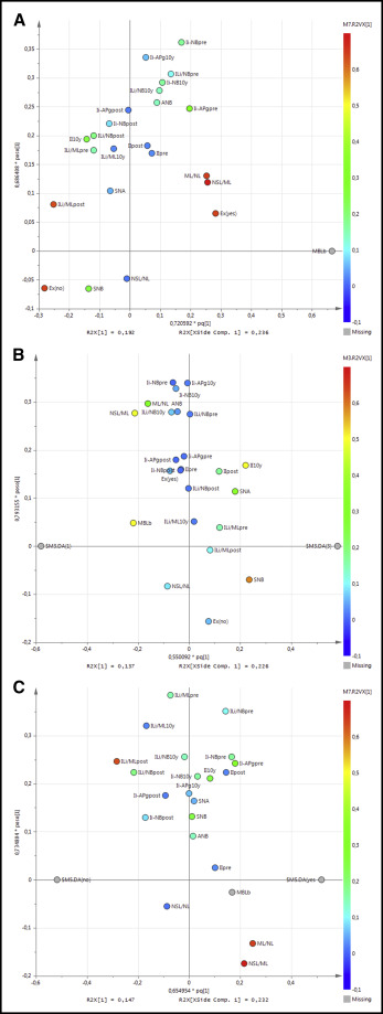
Multivariate data analysis (SIMCA version 13; Umetrics, Umeå, Sweden) was used to analyze and illustrate the differences between the groups and possible correlations between the primary and secondary variables. The orthogonal-PLS was used to illustrate possible correlations between the primary (y-variable) and secondary variables (x-variables). The orthogonal partial least squares projections to latent structures discriminant analysis was used to illustrate the differences between the groups.
Results
In the marginal bone levels, there were statistically significant differences between the groups for all surfaces, especially between the OR and C groups, but also between the O and C groups. Thus, the marginal bone levels were significantly lower for both the OR and O groups compared with the control group. Moreover, there was no significant difference between the OR and O groups regarding marginal bone level alterations ( Table II ; Fig 2 ).
| Variable | Retainer group (OR) | No retainer group (O) | Control group (C) |
|---|---|---|---|
| All surfaces | 11.3 (2.5) 11.5 (6.4; 16.5) |
11.0 (2.5) 10.2 (8.3; 15.9) |
8.74 (2.99) ∗ 8.35 (5.30; 15.57) |
| Buccal surface | 4.25 (1.36) † 4.04 (1.62; 6.28) |
3.88 (1.47) ‡ 3.58 (2.56; 8.23) |
2.57 (1.30) 2.47 (1.07; 5.29) |
| Lingual surface | 3.25 (1.33) 2.91 (1.69; 7.87) |
3.34 (1.38) 3.06 (1.86; 7.04) |
2.40 (1.01) 2.15 (1.18; 4.73) |
| Mesial surface | 1.98 (0.40) 1.92 (1.22; 2.80) |
1.98 (0.39) 1.90 (1.46; 2.84) |
2.02 (0.46) 1.90 (1.23; 2.93) |
| Distal surface | 1.83 (0.34) 1.81 (1.04; 2.46) |
1.84 (0.39) 1.87 (1.04; 2.70) |
1.74 (0.44) 1.72 (1.24; 2.62) |
A reduced marginal bone level on the buccal side of the mandibular front teeth was positively correlated with an open vertical basal relationship, a posterior rotation of the mandible (ML/NL, NSL/ML), extractions, protrusion (Ii-APg, and Ii-NB), and a high irregularity index at pretreatment. Furthermore, the reduced marginal bone level was negatively correlated with retroclined incisors (ILi/ML, ILi/NB) and a low irregularity index at posttreatment ( Fig 3 , A ).