The frequency and severity of untoward events associated with surgical procedures are influenced by multiple factors that may be related to the procedure, patient, and/or surgeon. Not every third molar needs to be removed. Full bony impacted lower third molars well below the cervical margin of the second molar crowns should be considered for retention. Certain deviations from normal healing should be considered to be complications. Risk factors associated with third molar removal should be carefully established and explained to the patient. Third molar surgery has a predictable postsoperative course for the average patient.
Complications are inevitably associated with the surgical management of third molars and invasive and noninvasive interventions in general. The frequency and severity of untoward events associated with surgical procedures are influenced by multiple factors that may be related to the procedure, patient, and/or surgeon ( Boxes 1–3 ). Complications associated with the removal of third molars and their management are discussed in this article.
-
Poor surgical access and visibility
-
Malfunctioning surgical drills or wrong anesthetic technique
-
Timing of surgical procedure
-
Aberrant tissue consistency
-
Wrong venue
-
Wrong or unnecessary procedure
-
Systemic comorbidities
-
Age of patient
-
Patient size
-
Unfavorable anesthetic candidate
-
Poor perioperative compliance
-
Limited space to deliver the luxated tooth
-
Large tongue
-
Small mouth
-
Limited jaw opening
-
Large, thick cheeks
-
Limited maxillary labial vestibule space
-
Errors in patient selection
-
Errors in choosing procedure
-
Poor surgical technique
-
Failure to honor patient’s complaints
-
Delay in identifying complications and introducing interventions
-
Unnecessary or imprudent surgery
-
Failure to thoroughly examine the patient clinically and radiographically
Perioperative anesthesia problems accompanying third molar surgery may or may not be directly related to tooth removal. Intraoral surgery, by its location and nature, poses a threat to the airway. Events related directly to the procedure, such as aspiration of a tooth fragment or a laryngospasm caused by poorly controlled irrigation fluids, can result in life-threatening consequences. Surgical misadventures are more likely to occur when the circumstances of an operation are adversely influenced by patient factors such as obesity, obstructive sleep apnea, large tongue, dense bone, and/or small mouth. Factors that contribute to poor visibility and accessibility increase the likelihood of surgical misadventures. A surgeon who is struggling to comfortably access a third molar site and persists in advancing the surgery increases the likelihood of an untoward surgical or anesthetic event. A fractious patient who is responding poorly to an office heavy sedation or general anesthetic introduces a cascade of negative influences that are likely to result in a surgical or anesthetic misadventure.
Not all third molar surgeries are alike. Not every third molar needs to be removed. Full bony impacted lower third molars that are well below the cervical margin of the second molar crowns should be considered for retention. Careful clinical examination should include periodontal probing of the distal surface of the second molar to rule out pocket depths of 4 mm or greater, which would indicate chronic periodontal inflammation .
Removal of lower third molars with dilacerated roots that extend below the inferior alveolar canal or into cortical bone at the inferior bone border with radiographic evidence of a disorder predicts inferior alveolar nerve damage and/or jaw fracture. Complications related to third molar removal are often related to errors of omission rather than commission. For example, complications are more likely to occur when the surgeon fails to perform an adequate preoperative patient evaluation and surgical site assessment. Careful patient selection and adherence to well-established indications for third molar removal reduce the more serious complications (lip and tongue numbness, fractured jaw) by, when indicated, choosing nonsurgical treatment.
This article provides an overview and listing of the common complications related to third molar surgery, their management, and prevention. Undesirable surgical outcomes may or may not be the result of poor surgeon judgment or technique. Complications are identified by their seriousness, reversibility, contributing factors, preventive measures, and management. Prudence and a noncavalier attitude toward third molar surgery are strongly encouraged.
Complications related to the procedure
▪
Intraoperative complications
In general, surgeons express the greatest concern about the more serious complications (broken jaw, numb lip/tongue, deep space infection) associated with third molar removal.
For the purposes of this analysis, in addition to the well-established intraoperative misadventures, any deviation from normal healing, including undue pain, swelling, and altered soft tissue and bony contour at the surgical site, is considered to be a complication.
-
Gingival clefting
-
Second molar loss of attached gingiva
-
Distal bone loss/periodontal pocket second molar
-
Persistent pain and tooth sensitivity
-
Temporomandibular disorder (TMD)/internal derangement
-
Lip lacerations
-
Corner-of-mouth abrasions
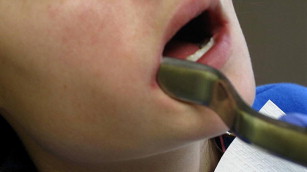
Reflecting a buccal mucogingival flap is usually the initial surgical intervention when removing impacted third molars. The flap should be carefully and delicately reflected to avoid tearing the margins of the gingival crevice and losing keratinized gingiva ( Fig. 1 ). Avoiding gingival clefting and loss of attached gingiva after third molar removal requires careful reflection of the buccal flap attached to the more anterior teeth. Torn buccal gingiva is more likely to occur when the interdental papillae are thin and minimally keratinized. Loss of superior buccal plate and periodontal pocket defects with associated periodontal inflammation increase the likelihood of injuring the gingiva. Immediate repair may reduce the prospect of long-term gingival contour loss. Vertical releasing incisions should be precisely sutured and the envelope flap positioned snuggly to the cervical margins of the more anterior teeth.
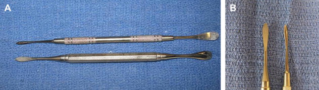
Dental papillae should be delicately reflected, avoiding crushing or tearing. Failure to maintain flap integrity delays healing, invites greater swelling and pain, results in loss of gingival contour and quality (keratinized gingiva), and contributes to gingival clefts and chronic irritation. Sharply incising into the gingival crevice and using a pointed instrument such as a wax spatula rather than a molt 9 periosteal elevator facilitates uncovering the third molar surgical site ( Fig. 2 ).
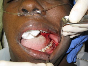
Visibility and accessibility of the impacted tooth determine the extent and mobility of the flap to avoid tearing and compromised surgical access. The back edge of the scalpel blade can lacerate the lower lip when exposing maxillary third molars in patients with limited surgical access, or can the mouth commissures can be abraded with retractors ( Fig. 3 ). Damaged retractors or suction tips can cause cheek and mouth floor soft tissue injuries while exposing the surgical field and keeping it dry. Loss of gingival contour or substance may require gingival grafting ( Fig. 4 ).
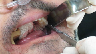
Distal bone loss at the second molar site and periodontal pocket inflammation following third molar removal are more likely to occur when a fully developed lower third molar crown is located below the cervical margin of the second molar in intimate proximity to the root structure of the erupted molar. Intraoperative inspection of the distal surfaces of the second molar should be routinely enacted. Wide exposure of the distal root surface of the second molar is an indication for immediate socket preservation. Loss of soft tissue and bone at the distal margin of the second molar invites pain and tooth sensitivity. Defects on the distal margin of the second molar after lower third molar removal are less likely when the third molar’s crown and cervical root third are developed .
Intraoperative complications
In general, surgeons express the greatest concern about the more serious complications (broken jaw, numb lip/tongue, deep space infection) associated with third molar removal.
For the purposes of this analysis, in addition to the well-established intraoperative misadventures, any deviation from normal healing, including undue pain, swelling, and altered soft tissue and bony contour at the surgical site, is considered to be a complication.
-
Gingival clefting
-
Second molar loss of attached gingiva
-
Distal bone loss/periodontal pocket second molar
-
Persistent pain and tooth sensitivity
-
Temporomandibular disorder (TMD)/internal derangement
-
Lip lacerations
-
Corner-of-mouth abrasions
Reflecting a buccal mucogingival flap is usually the initial surgical intervention when removing impacted third molars. The flap should be carefully and delicately reflected to avoid tearing the margins of the gingival crevice and losing keratinized gingiva ( Fig. 1 ). Avoiding gingival clefting and loss of attached gingiva after third molar removal requires careful reflection of the buccal flap attached to the more anterior teeth. Torn buccal gingiva is more likely to occur when the interdental papillae are thin and minimally keratinized. Loss of superior buccal plate and periodontal pocket defects with associated periodontal inflammation increase the likelihood of injuring the gingiva. Immediate repair may reduce the prospect of long-term gingival contour loss. Vertical releasing incisions should be precisely sutured and the envelope flap positioned snuggly to the cervical margins of the more anterior teeth.
Dental papillae should be delicately reflected, avoiding crushing or tearing. Failure to maintain flap integrity delays healing, invites greater swelling and pain, results in loss of gingival contour and quality (keratinized gingiva), and contributes to gingival clefts and chronic irritation. Sharply incising into the gingival crevice and using a pointed instrument such as a wax spatula rather than a molt 9 periosteal elevator facilitates uncovering the third molar surgical site ( Fig. 2 ).
Visibility and accessibility of the impacted tooth determine the extent and mobility of the flap to avoid tearing and compromised surgical access. The back edge of the scalpel blade can lacerate the lower lip when exposing maxillary third molars in patients with limited surgical access, or can the mouth commissures can be abraded with retractors ( Fig. 3 ). Damaged retractors or suction tips can cause cheek and mouth floor soft tissue injuries while exposing the surgical field and keeping it dry. Loss of gingival contour or substance may require gingival grafting ( Fig. 4 ).
Distal bone loss at the second molar site and periodontal pocket inflammation following third molar removal are more likely to occur when a fully developed lower third molar crown is located below the cervical margin of the second molar in intimate proximity to the root structure of the erupted molar. Intraoperative inspection of the distal surfaces of the second molar should be routinely enacted. Wide exposure of the distal root surface of the second molar is an indication for immediate socket preservation. Loss of soft tissue and bone at the distal margin of the second molar invites pain and tooth sensitivity. Defects on the distal margin of the second molar after lower third molar removal are less likely when the third molar’s crown and cervical root third are developed .
Complications related to the surgeon
Injudicious extraction forces, excessive opening of the mouth with props, and preexisting internal derangements of the temporomandibular junction invite post–third molar TMD. Surgeon and assistant stabilization of the mandible when forceps and elevator forces are being exerted is necessary. Mouth opening should not exceed the patient’s comfortable interincisal distance.
Abrasions, lacerations, and burns to the lip and corner of the mouth can be extensive enough to cause permanent skin and/or commissure scarring. Removing superiorly positioned impacted upper third molars under conditions of limited access and visibility invites soft tissue injury ( Fig. 5 ). An overheated handpiece can cause substantial scarring and lip disfigurement. Marred retractors and suction tips made sharp and irregular during previous surgeries should be screened for and replaced.
-
Fractured mandible
-
Numb lip
-
Numb tongue
-
Displacement of upper third into the infratemporal fossa
-
Displacement of upper third into the maxillary sinus
Risk factors for mandible fracture
Fractures of the mandibular angle associated with third molar removal are generally avoidable when precise preoperative clinical and radiographic evaluations are completed. Factors that increase the risk of jaw fracture are shown in Box 4 . The more risk factors present, the more likely a mandibular angle fracture will occur. When the risk of fracture is high, the indications for removal of a lower third molar should be compelling. Preoperative discussions informing the patient of the increased risk of a broken jaw are critical. Performing the surgical procedure in the hospital operating room (OR) with the patient nasally intubated allows the surgeon to concentrate on the third molar removal, and this is the best venue to immediately repair the fracture should one occur. Judicious bone removal and applying modest elevator forces to the carefully sectioned tooth are vital. Removing less bone and sectioning the tooth into multiple pieces may be a reasonable surgical approach. Root tips lodged in the dense cortical inferior mandibular bone or in the inferior alveolar canal may be best left in place. Application of arch bars and maxillomandibular fixation may be prudent when the mandible is still intact after third molar removal but is at risk for postoperative fracture under normal chewing function and physical activities ( Figs. 6–8 ).
-
Older patient
-
Dilacerated and flared roots
-
Dense bone
-
Metabolic bone disease
-
Osteomyelitis
-
Small mandible
-
Third molar occupies the height of the jaw
-
Associated odontogenic disorders
-
Injudicious surgical forces
Stay updated, free dental videos. Join our Telegram channel
VIDEdental - Online dental courses


