LeFort I osteotomy, anterior segmental osteotomy, bilateral sagittal split ramus osteotomy, and genioplasty are frequently used methods for correcting facial deformities. However, in patients with an abnormally shaped maxilla or mandible, more complex surgical techniques or multiple combinations must be considered for improved esthetic results. This article presents a patient with bialveolar protrusion, mandibular prognathism, chin retrusion, a long face, and severe facial asymmetry. A combination of LeFort I asymmetric impaction, anterior segmental osteotomy, and 3-piece segmentation of the maxilla, and bilateral sagittal split ramus osteotomy, anterior segmental osteotomy, genioplasty advancement, and angle shaving in the mandible were conducted simultaneously. In patients with complicated deformities that cannot be classified by simple conventional classification methods, multisegmental osteotomy can be an option for improved esthetic results.
Among the various surgical techniques for correcting a facial deformity, LeFort I osteotomy, anterior segmental osteotomy, bilateral sagittal split ramus osteotomy, and genioplasty might be the most frequently performed. However, for patients with an abnormally shaped maxilla or mandible, more complex surgical techniques or a combination of multiple surgeries should be considered for improved esthetic results. This approach requires excellent surgical skills and sufficient bone volume but can also induce many problems: eg, aseptic necrosis of segments, increased bleeding, problems of wound healing, tooth pulp damage, root injury, and periodontal problems. As surgical skills and knowledge develop, however, the complications have decreased. Many authors claim that, with basic surgical principles and a meticulous treatment plan, multisegmentation surgery is a safe option associated with only limited and minor complications.
We present a patient with bialveolar protrusion, mandibular prognathism, chin retrusion, a long face, and severe facial asymmetry. To correct these problems, orthodontic retraction of the maxillary anterior teeth with premolar extraction and conventional 2-jaw surgery including LeFort I osteotomy, bilateral sagittal split ramus osteotomy setback, and genioplasty advancement were options. This treatment, however, would have resulted in insufficient correction of the bialveolar protrusion and chin retrusion. Therefore, a combination of LeFort I asymmetric impaction, anterior segmental osteotomy, and segmentation of the maxilla, and bilateral sagittal split ramus osteotomy, anterior segmental osteotomy, genioplasty advancement, and angle shaving in the mandible were performed.
Diagnosis
A 30-year-old woman with chief complaints of anterior protrusion, chin retrusion, and facial asymmetry visited the clinic of the first author (U.-B.B.). She had a convex profile with protruding lips and showed lip incompetency with hyperactive mentalis activity ( Figs 1 and 2 ). The mandible was prognathic, but the chin was retruded ( Fig 3 ). The numeric values of her cephalometric skeletal classification such as ANB and SNA carried less meaning than usual because she had a deformation of the jaw bone itself. The problem was that a setback of the mandible for correction of the anterior protrusion could exacerbate her chin retrusion.
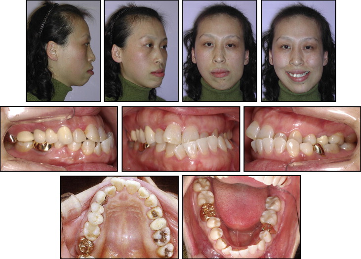
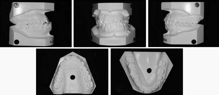
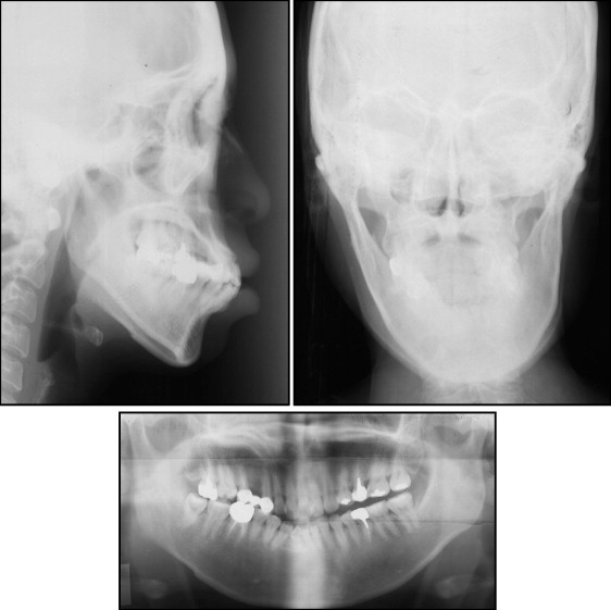
The patient also had severe facial asymmetry. In the maxilla, there was occlusal plane canting, with the right side tipped downward by 3 mm. The mandible was deviated to the left side. A severe difference in the right and left gonial angle areas was also obvious. The maxillary dental midline was deviated 2.0 mm to the right side, and the mandibular dental midline was deviated 4.0 mm to the left. In a coronal view, yawing of the entire maxilla to the right side was also observed. She also had a long face, a gummy smile, and vertical maxillary excess.
In the dental analysis, the canines and molars showed Class III relationships on the right side, with a Class I molar relationship on the left side. These relationships were mainly due to the left shift of the mandible. A buccal crossbite was noted on the left side, and a collapsed bite was present. A missing maxillary right first premolar had been replaced with a 3-unit porcelain fused to metal bridge.
In the temporomandibular joint evaluation, the right condyle was longer than the left one. However, she experienced no specific symptoms such as clicking, pain, or mouth-opening problems.
Treatment objectives
The treatment objectives were as follows.
- 1.
Sagittal: correct the bialveolar protrusion and chin retrusion.
- 2.
Coronal: correct the maxillary canting, yawing, and mandibular asymmetry.
- 3.
Vertical: correct the gummy smile and long face.
- 4.
Dental: achieve Class I molar and canine relationships, flatten the occlusal plane, and coordinate the arch widths.
Treatment alternatives
Orthodontic retraction of the maxillary anterior teeth with premolar extraction and conventional 2-jaw surgery, such as a LeFort I osteotomy, bilateral sagittal split ramus osteotomy setback, and genioplasty advancement, was a treatment option. This method, however, would result in insufficient correction of the bialveolar protrusion. Therefore, anterior segmental osteotomy was chosen instead to retract the anterior teeth and the alveolar bone together to a greater amount.
There was a crossbite on the left side except for the left first and second molars. After the bilateral sagittal split ramus osteotomy and the asymmetric setback of the mandible, the maxillary arch became wider than the mandibular arch. To correct these problems, both orthodontic and surgical approaches were possible. Orthodontic constriction, such as a quad helix or a precision lingual arch, had the advantage of reducing surgical trauma but also had disadvantages of difficulty in torque control and lengthened treatment time.
Surgical constriction such as midline osteotomy and a Y-shaped 3-piece osteotomy were other options. In this patient, anterior segmental osteotomy could result in steps or arch discontinuity between the canine and the second premolar. Both to constrict the maxillary arch and to reduce arch discontinuity after the anterior segmental osteotomy, a Y-shaped 3-piece osteotomy was chosen. As a result, in the maxilla, LeFort I osteotomy asymmetric impaction with a 3-piece Y-shaped osteotomy including an anterior segmental osteotomy setback was planned.
It was necessary to set back the mandible surgically to correct the anterior crossbite and the mandibular prognathism. However, for this patient, such a procedure could have aggravated the chin retrusion. The direction and the degree of deformity of the alveolar bone were not aligned with those of the basal bone. The main treatment objective in the sagittal plane was posterior movement of the mandibular anterior proclination with simultaneous anterior movement of the chin. Therefore, the combination of anterior segmental osteotomy setback and advancement genioplasty was chosen for the mandible.
Treatment progress
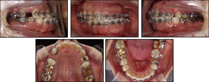
After cutting the 3-unit-bridge and removing the pontic, fixed 0.018 × 0.025-in preadjusted appliances were placed. Through leveling and alignment, the mandibular anterior teeth proclined labially; this relieved the deep overbite and flattened the occlusal plane ( Fig 4 ). This process took 16 months ( Figs 5 and 6 ). Before the surgery, segmented archwires were inserted from canine to canine and from each second premolar to each second molar in the maxillary and mandibular arches for the anterior segmental osteotomy and the first premolar extractions.
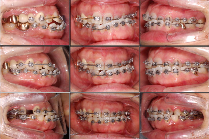
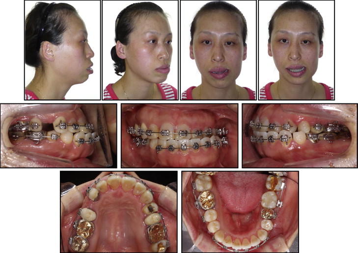
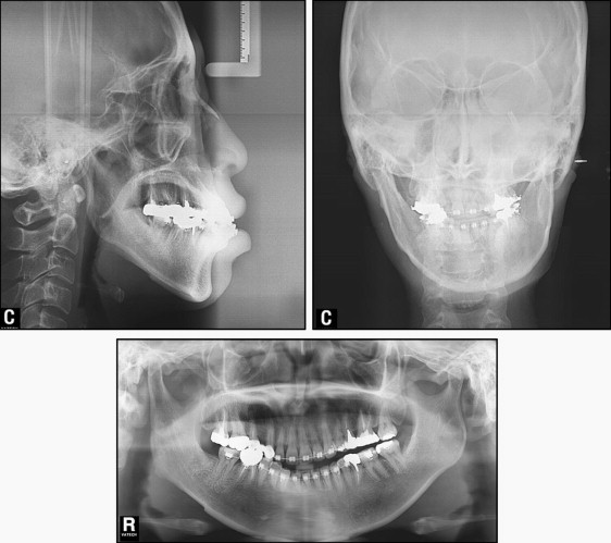
Surgery was performed with the extraction of 4 first premolars. In the maxilla, a LeFort I osteotomy was carried out with a Y-shaped, 3-piece osteotomy.
- 1.
LeFort I osteotomy was performed. The maxillary midline moved 2.0 mm to the left. The maxilla was impacted differently on the right and left sides, with impactions of 6 mm on the right side and 3 mm on the left side. These procedures corrected the yawing and occlusal plane canting of the maxilla.
- 2.
Arch width was reduced by 3 mm, with simultaneous torque control of the posterior alveolar segments.
- 3.
Anterior segmental osteotomy was conducted with extraction of the first premolars. A setback of the anterior segment by 5 mm was performed with simultaneous linguoversion for adequate incisor torque. The posterior segments were advanced by 1.5 mm, and the remaining space was left to be treated during the postoperative orthodontic therapy. In the mandible, bilateral sagittal split ramus osteotomy, anterior segmental osteotomy, genioplasty, and angle reduction were performed.
- 4.
Bilateral sagittal split ramus osteotomy was performed to correct the mandibular asymmetry. Through differential setbacks of the right and left distal segments (8 mm on the right and 2 mm on the left), the chin deviation was corrected.
- 5.
Anterior segmental osteotomy with extraction of the first premolars was carried out to correct the mandibular anterior alveolar protrusion. The anterior alveolar bone of the mandible was moved backward by this procedure.
- 6.
Advancement genioplasty by 5 mm with height reduction of 2 mm was achieved.
- 7.
Angle reduction was performed to correct the asymmetric gonial angles.
Through the postoperative orthodontic treatment, the occlusal steps between the anterior and posterior segments of the anterior segmental osteotomy were leveled and aligned ( Fig 7 ). The remaining spaces were closed. During space closure, an anterior edge-to-edge occlusion appeared. Screws were inserted in the mandibular posterior region to retract the mandibular incisors with maximum anchorage.

