Case• 4. Gingival recession
SUMMARY
A 30-year-old woman has gingival recession. Assess her condition and discuss treatment options.
History
Complaint
The patient is worried about the gingival recession around her lower front teeth, which she feels is worsening.
History of complaint
She remembers noticing the recession for at least the previous 5 years. She thinks it has worsened over the last 12 months. There has recently been some sensitivity to hot and cold and gingival soreness, most noticeably on toothbrushing or eating ice cream.
Dental history
The patient has been a patient of your practice for about 10 years and you have discussed her recession at previous visits and reassured her. She has a low caries rate and generally good oral hygiene.
Medical history
She is a fit and healthy individual and is not a smoker.
▪ What further specific questions would you ask to help identify a possible cause?
How often do you brush your teeth? Provided brushing is effective, cleaning once a day is sufficient to maintain gingival health. However, most patients clean two or three times each day and some brush excessively in terms of frequency, duration and force used. Trauma from brushing is considered a factor in some patients’ recession, and recession may indicate a need to reduce the frequency and duration of cleaning while maintaining its effectiveness. In this instance the patient has a normal toothbrushing habit but should clean no more than twice each day and for a sensible period of time.
Have you had orthodontic treatment? A lower incisor is missing, suggesting that some intervention may have taken place. Fixed orthodontics in the lower labial segment is occasionally associated with gingival recession in patients with thin buccal gingiva, narrow alveolar processes and correction of severe crowding. Plaque control may be compromised during the wearing of an orthodontic appliance and, even over a relatively short period, this can contribute to the problem. In this instance the patient had undergone extraction of the incisor but had not worn an appliance.
Examination
Intraoral examination
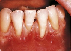
▪ The appearance of the lower incisors is shown inFigure 4.1. What do you see?
— Missing lower left central incisor.
— Unrestored teeth.
— No plaque is visible except for a small amount at the cervical margin of the lower left lateral incisor.
— Gingival recession affecting all lower incisors and, to a lesser extent, the lower canines.
— Apart from the abnormal contour, the buccal gingivae are pink and healthy and the interdental papillae are normal.
— Reduction in width of keratinized (cornified) attached gingival epithelium. In places, attached gingiva appears absent.
▪ What clinical assessments would you make, how would you make them and why are they important?
See Table 4.1.
| Assessment | Method | Importance |
|---|---|---|
| Recession | Measure from the gingival margin to the cement–enamel junction | Provides baseline readings to assess progression |
| Probing depths | Routine periodontal probing | Detects associated loss of attachment undermining the reduced width of attached gingiva |
| Bleeding on probing | Routine recording of bleeding on probing; immediate or delayed | Indicates the presence of gingival inflammation and poor oral hygiene |
Stay updated, free dental videos. Join our Telegram channel
VIDEdental - Online dental courses


