Introduction
The aims of this study were to assess the biologic stability of a newly designed hollow (H-type) miniscrew compared with conventional (C-type) miniscrews through histomorphometric and histologic analysis.
Methods
Both types of miniscrews were placed into the maxillae and the mandibles of 12 beagles. Maximum insertion torque, Periotest (Siemens AG, Bensheim, Germany) value, bone-implant contact, and bone volume were measured.
Results
The overall success rates of the H-type were 78.3% in the maxilla and 60.0% in the mandible. Mean maximum insertion torque values of the H-type were 14.2 N-cm in the maxilla and 20.9 N-cm in the mandible. The Periotest values of the H-type were −1.5 in the maxilla and −6.4 in the mandible. Mean maximum insertion torque and Periotest values of the H-type were higher than those of the C-type. In the maxilla, the bone-implant contact values of the H-type were 37.3% and 32.3% at 3 and 12 weeks, respectively. In the mandible, the bone-implant contact values were 31.4% and 18.5% at 3 and 12 weeks, respectively.
Conclusions
Considering the lower success rate and the insufficient bone-implant contact and bone volume of the H-type in the mandible, the clinician should choose a suitable combination of miniscrews depending on local bone quality and implantation site, such as an H-type in the maxilla and a C-type in the mandible.
The development of temporary anchorage devices such as miniscrews, palatal implants, retromolar implants, and bone plates has provided maximum anchorage in the biomechanics of orthodontics. Miniscrews have been used most often for absolute anchorage; their versatility has aroused interest in their primary stability and their failure.
The primary stability of miniscrews is mainly supported by mechanical retention of the bone-to-implant interface. Miniscrew design, screw diameter, and loading protocols affect primary stability, as does bone quality, especially the thickness of cortical bone. Bone density and the cortical-to-cancellous ratio of bone certainly influence miniscrew stability at placement.
Recently, various miniscrew designs have been developed to improve the primary stability. A larger-diameter miniscrew showed a significant increase in insertion torque, which agreed with a finite element method study of stress distribution over the length and diameter of the orthodontic miniscrews. In addition, a tapered miniscrew displayed maximum insertion torque compared with a cylindrical miniscrew. However, the length of the miniscrews had no effect on their survival.
One method for assessing miniscrew stability is to measure insertion torque. However, high insertion torque does not necessarily result in stronger fixation. Overtightening can lead to continuous compression of the surrounding bone and thus cause microfracturing of the bone threads around the miniscrew. Degeneration of the bone at the interface might aggravate bone regeneration surrounding the miniscrew thread.
In addition to insertion torque, bone-implant contact (BIC) is essential for fixation strength in primary stability and for osseointegration in secondary stability. The Periotest (Siemens AG, Bensheim, Germany) can be easily used as a nondestructive method for measuring miniscrew stability.
Primary stability is an essential prerequisite for the success of miniscrews, but other factors can affect success rates. Since root contact is a major factor in miniscrew failure in orthodontic anchorage, a hollow-centered miniscrew (H-type) was developed. This miniscrew is wide enough to improve stability and short enough to allow placement in the bone superficial to the root surface as well as intraradicular bone; its tapered design can enhance initial fixation. Furthermore, this new miniscrew has a hollow interior to promote insertion and to compensate for the relatively lower amount of bone contact because of its shorter length, with bone formation occurring in the interior part of the miniscrew. This miniscrew also contains 4 fenestration holes in the beginning of the first thread for ingrowth of bone.
Hong et al previously investigated the mechanical stability of the H-type of miniscrew implant inserted into biosynthetic bone blocks and assessed this stability with cone-beam computed tomography. However, these studies lacked histologic analyses that examined bone remodeling and cellular responses around the miniscrews, and they did not evaluate BIC and bone volume (BV) in the interior of the miniscrews.
The aim of this animal-based study was to assess the biologic stability of the H-type of miniscrew. We evaluated insertion torque, BIC, and BV around the miniscrew in beagles and compared the biologic stability of H-type miniscrews with conventional (C-type) miniscrews. In addition, we used histology to observe bone remodeling around the bone-to-miniscrew interface and the interior part of the H-type of miniscrew.
Material and methods
Twelve adult male beagles (age, 12-15 months; weight, 10-14 kg) were used in this study and maintained in the animal laboratory at Yonsei University Medical Research Center in Seoul, Korea; the experimental protocol was approved by the institutional animal care and use committee.
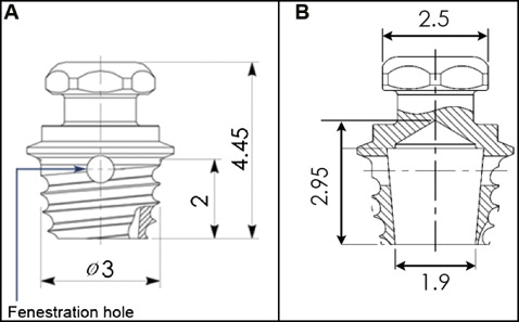
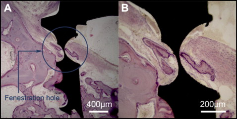
The 12 dogs were divided into C-type and H-type miniscrew groups, with a split-mouth design. Thirty-eight self-drilling H-type miniscrews (diameter, 3.0 mm; length, 2.0 mm; tapered shape; Biomaterials Korea, Seoul, Korea) were placed into the intraradicular spaces (just below the furcation area) of the beagles’ maxillary and mandibular first molars and the mandibular fourth premolars ( Fig 1 ). The H-type, single-threaded, tapered miniscrew is a hollow miniscrew with 4 fenestration holes in the beginning of the first thread for the ingrowth of bone ( Fig 2 ).
Thirty-seven self-drilling C-type miniscrews (diameter, 1.45 mm; length, 7 mm; single-threaded, cylindrical shape; Biomaterials Korea) were placed around the second and third premolars in the maxilla and the mandible.
All experimental procedures, including surgical procedures and clinical examinations such as taking intraoral photographs, were performed aseptically. The experimental animals were injected subcutaneously with 0.02 mg per kilogram of atropine, intramuscularly with 5 mg per kilogram of enrofloxacine, intravenously with 0.5 mg per kilogram of ketorolac tromethamine, and intravenously with 5 mg per kilogram of cimetidine (premedication), followed by zoletil (5 mg/kg body weight; Zoletil 50; Virbac Korea, Seoul, Korea) and xylazine (0.2-0.5 mg/kg body weight; Rompun; Bayer, Leverkusen, Germany) intravenously to induce general anesthesia. Throughout the operation, 2% enflurane or 1% to 2% isoflurane was injected for maintenance.
The beagles that received C-type and H-type miniscrews were further divided into 2 groups to examine 3-week and 12-week experimental periods.
The safe zones for miniscrew insertion in beagles have been reported to be located in the intraradicular space of the first molar, the intraradicular space of the fourth premolar, and the interradicular space between the fourth premolar and the first molar in the mandible. In the maxilla, the intraradicular space of the first molar and the intraradicular space of the second and third premolars are safe zones for miniscrew insertion ( Fig 3 , A ). The locations for miniscrew placement were determined from these studies.
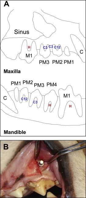
The implantation procedure was performed under saline-solution irrigation without flap surgery, with only a 5-mm gingival incision in the implantation site to prevent tissue impingement. On average, the distance between 2 adjacent miniscrews was 1.1 to 1.5 mm. All miniscrews were inserted perpendicular to the bone surface until their necks contacted the bone ( Fig 3 , B ).
During the entire experimental procedure, chlorhexidine solution was applied daily to maintain oral hygiene.
The first miniscrews were inserted 12 weeks before the dogs were killed; after 9 weeks, a second miniscrew was inserted ( Fig 4 ). After 3 more weeks, all experimental animals were killed.

All miniscrews in the C-type and H-type groups were confirmed as fully inserted by checking the bone contact of the final screw thread with a dental explorer. The highest insertion torque was measured in Newton centimeters during an initial quarter turn using a torque sensor (MGT50; Mark-10, New York, NY). Initial screw mobility was measured twice on each miniscrew with the Periotest after insertion. The mean value of the 2 measurements was recorded as the initial mobility.
After 12 weeks, all experimental animals were killed, and tissue blocks containing the miniscrews and adjacent teeth were prepared. Tissue blocks were fixed in 10% formalin for 1 month. After fixation, the tissues were dehydrated in progressively higher concentrations of alcohol (70%-100%) for 2 weeks. The dehydrated, fixed tissues were embedded in Technovit 8000 (Heraeus Kulzer, Wehrheim, Germany), cut with a diamond saw parallel to the miniscrew axis, and ground into serial sections (approximately 20 μm) using the Exakt grinding system (Exakt Technologies, Oklahoma City, Okla). Hematoxylin and eosin staining was performed for histologic observations and histomorphometric analyses.
Optical microscopic images were taken with a DM LB microscope (Leica, Wetzlar, Germany), and histomorphometric parameters were measured at 100 times magnification. Tissue specimens were examined on the microscope and analyzed with an image analysis program linked to a light microscope using IMT iSolution Lite (version 8.1; IMT iSolution, Jena, Germany) and Image-Pro Plus (version 4.5.0.29; Media Cybernetics, Silver Spring, Md).
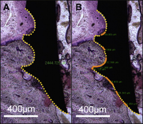
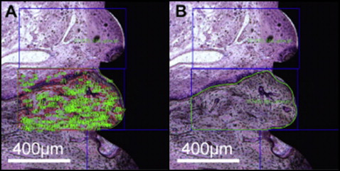
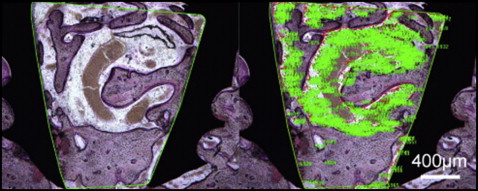
Histomorphometric analysis was performed in the regions of interest, within 800 μm of the implant-bone interface ( Fig 5 ). BIC and the ratio of BV to total volume (BV/TV) were calculated as percentages from the regions of interest. In addition, histologic analyses were performed to observe bone remodeling around the bone-to-miniscrew interface and around the interior part of the H-type miniscrew.
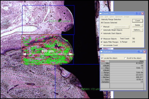
BIC (%) was calculated as the length of the BIC divided by the total length of the implant interface, multiplied by 100%. BV/TV (%) was calculated as BV divided by TV, multiplied by 100%. For the H-type miniscrews, BIC and BV/TV were measured in both their outer surfaces ( Figs 6 and 7 , respectively) and the inside of the hollow portion of the miniscrews ( Fig 8 ).
Statistical analysis
SPSS software (version 17.0; SPSS, Chicago, Ill) was used for statistical analysis. The success rates of the C-type and H-type miniscrews were calculated in the maxilla and the mandible. For the Periotest value and the insertion torque, the means and standard deviations were calculated. Two-way analysis of variance (ANOVA) was used to compare mean insertion torque or mean Periotest value according to group (C-type and H-type) and jaw (maxilla and mandible). For BIC and BV/TV, the means and standard deviations were calculated, and 2-way ANOVA was used to compare BIC and BV/TV according to jaw and experimental period (3 and 12 weeks). The BIC and BV/TV values for the exterior and interior parts of the H-type miniscrews were compared with 2-way ANOVA according to jaw and experimental period. P <0.05 was considered to indicate statistical significance.
Results
The failure criterion was defined as “fell out of the alveolar bone.” In the H-type group, 5 of 23 miniscrews in the maxilla and 6 of 15 miniscrews in the mandible fell out. The success rates were 78.3% in the maxilla and 60.0% in the mandible in the H-type group. The success rate in the C-type group was 100% in both the maxilla and the mandible.
The mean insertion torque values in the H-type group were 14.2 N-cm in the maxilla and 20.9 N-cm in the mandible ( Table I ). Insertion torque significantly differed between the C-type and H-type groups ( P <0.0001); measurements in the H-type group were significantly higher than those in the C-type group in both the maxilla and the mandible. Furthermore, the insertion torque was significantly higher in the mandible than in the maxilla in both groups. There was no interaction between groups and jaws with 2-way ANOVA ( P = 0.2101).
| Jaw | Insertion torque (N-cm) | Significance | |||
|---|---|---|---|---|---|
| C-type | H-type | ||||
| Mean | SD | Mean | SD | ||
| Maxilla | 8.43 | 3.40 | 14.18 | 5.05 | † |
| Mandible | 11.88 | 5.12 | 20.86 | 3.08 | ‡ |
| Significance | ∗ | † | |||
The mean Periotest value was significantly lower in the H-type group, with a significant difference in the mandible ( P <0.0001). The mean Periotest value in the C-type group was significantly lower in the maxilla, and the mean Periotest value in the H-type group was significantly lower in the mandible ( Table II ).
| Jaw | Periotest value | Significance | |||
|---|---|---|---|---|---|
| C-type | H-type | ||||
| Mean | SD | Mean | SD | ||
| Maxilla | −0.11 | 2.69 | −1.49 | 2.74 | NS |
| Mandible | 2.85 | 3.13 | −6.42 | 0.79 | † |
| Significance | ∗ | † | |||
The mean BIC in both groups declined in the maxilla and the mandible over the experimental period, but there was no significant difference between time points. In the mandibles, there was a significant difference between the groups at 12 weeks ( Table III ). The mean BV/TV in both groups declined over the experimental period. Mean BV/TV significantly decreased over the experimental period in the mandibles of the H-type group ( Table III ). The mean BIC in the C-type group was significantly higher in the mandible ( P <0.05) than in the maxilla ( Table IV ).
| Jaw | Time (wk) | BIC (%) | Sig | BV/TV (%) | Sig | ||||||
|---|---|---|---|---|---|---|---|---|---|---|---|
| C-type | H-type | C-type | H-type | ||||||||
| Mean | SD | Mean | SD | Mean | SD | Mean | SD | ||||
| Maxilla | 3 | 30.19 | 12.02 | 37.29 | 21.86 | NS | 62.75 | 15.57 | 47.34 | 19.74 | NS |
| 12 | 21.50 | 8.28 | 32.31 | 23.33 | NS | 44.28 | 8.57 | 40.45 | 11.82 | NS | |
| Sig | NS | NS | NS | NS | |||||||
| Mandible | 3 | 48.65 | 15.38 | 31.36 | 17.81 | NS | 49.59 | 11.41 | 51.84 | 25.58 | NS |
| 12 | 45.28 | 12.53 | 18.45 | 21.43 | ∗ | 48.89 | 13.33 | 37.45 | 30.74 | NS | |
| Sig | NS | NS | NS | ∗ | |||||||
| Time (wk) | Jaw | BIC (%) | Sig | BV/TV (%) | Sig | ||||||
|---|---|---|---|---|---|---|---|---|---|---|---|
| C-type | H-type | C-type | H-type | ||||||||
| Mean | SD | Mean | SD | Mean | SD | Mean | SD | ||||
| 3 | Mx | 30.19 | 12.02 | 37.29 | 21.86 | NS | 62.75 | 15.57 | 47.34 | 19.74 | NS |
| Mn | 48.65 | 15.38 | 31.36 | 17.81 | NS | 49.59 | 11.41 | 51.84 | 25.58 | NS | |
| Sig | ∗ | NS | NS | NS | |||||||
| 12 | Mx | 21.50 | 8.28 | 32.31 | 17.81 | NS | 51.96 | 15.58 | 40.45 | 11.82 | NS |
| Mn | 45.28 | 12.53 | 18.45 | 21.43 | ∗ | 48.89 | 13.33 | 37.45 | 30.74 | NS | |
| Sig | ∗ | NS | NS | NS | |||||||
Stay updated, free dental videos. Join our Telegram channel

VIDEdental - Online dental courses


