We agree that the term “hybrid” is appropriate if the dentition is used with the implants for anchorage for the expansion force. However, this does not mean that incorporating the dentition in an appliance design always makes it a hybrid or dental expander. If the purpose of the dentition in the appliance is not anchorage for the expansion force but something else, then the term “true skeletal anchorage” can be applied to the appliance. The expander we used, which we now officially call the “maxillary skeletal expander” (MSE), was originally designed to deliver the expansion force to 4 implants inserted deeply, engaging both layers of the cortical bone (palatal and nasal layers); the first molars were used to stabilize the position of the jackscrew during the expansion rather than for anchorage. The interlocked suture in mature patients undergoes significant torsion in 3 dimensions as it splits, and the 4 implants will undergo unnecessary strain and tip as the 2 halves of the maxilla twist away from each other. Using the maxillary molars to stabilize the jackscrew in 3-dimensional space during the expansion reduces this unwanted strain on implants, preventing breakage and loosening. Thus, this appliance can be categorized as a “skeletal anchorage device” as long as the implants are solely subjected to expansion force.
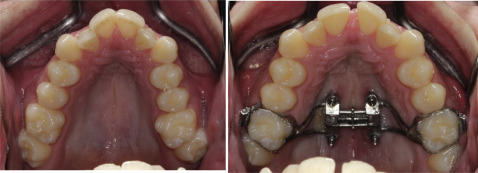
However, the appliance can become a hybrid if the implants fail or tip and the expansion force transfers to the molars. This can be evaluated easily by comparing the buccal positions of the dentition in contact with the MARPE against the ones not in contact. Figure 1 shows that an MSE can cause skeletal expansion without dental movements. If the expansion force is directed at first molars that are connected to the MSE, the molars would move laterally, leaving the adjacent teeth behind, since the other teeth are not in contact with the appliance. However, the relationship between the first molars and adjacent teeth did not change, indicating that the expansion was skeletal. The MARPE used in our report was a modified version of the MSE, with palatal wire extensions, making the evaluation more difficult. Figure 2 shows occlusal photographs of our patient before and after the suture split. Even here, the relative buccal positions of the first and second molars did not change. The first molars were connected to the MARPE, but the second molars were not in contact with the appliance. The right second premolar was not in contact with the palatal extension, but its relative position did not change either, indicating that the skeletal expansion occurred with no evidence of dental movement. The dental movements observed in the final records in our case report most likely occurred during the orthodontic phase of treatment, after the removal of this appliance.


