First, we thank the authors for their interest in our article and the comments and matters raised. However, we think that some points need to be clarified.
The stable reference landmarks used in our study were the lateral and medial points of the third rugae area as recommended in previous studies. The blue and green areas in Figure 4, C , show significantly more surface area than the red color, especially toward the medial and lateral parts of the third rugae area that are the most stable points. To make sure that we reproduced the same measurements, we have measured the reliability coefficient (Cronbach alpha) of the randomly collected data (from six 3-dimensional study models with a 2-week interval). The reliability coefficient was 0.95, which suggested accurate superimpositions. Moreover, the color-matching level was minor compared with other studies that reflected the accuracy of the software used. For instance, in our study, the mismatched color levels were 0.00-0.02, 0.02-0.05, 0.05-0.08, and 0.08-0.1 mm (Fig 4, C ); in other studies, the mismatched levels were 0.00-0.4, 0.5-1.0, 1.0-1.4, and 1.4-1.6 mm for blue, green, yellow, and red, respectively.
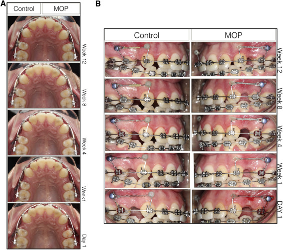
The images presented in our study in Figures 4 and 5 were used for demonstration, and this was model number 28 between the second and third months’ digital models. Hence, it showed already retracted canines. However, here we present the same subject at the baseline, and the first, second, and third months to show the validity of our records without any doubts ( Fig 1 ). We felt that it was inappropriate to judge and invalidate our study based on 1 figure that we included to illustrate the technique. This study was a randomized controlled trial with a decent sample size and standardized precise measurement methods analyzed to show the conclusive results by numbers, not by eye polling. For readers who like to see pictures, Figures 1 and 2 in our reply show 2 different cases Figure 2 shows more canine displacement on the MOP side (week 8), and Figure 3 (weeks 8 and 12) shows more canine retraction on the control side. However, the same amounts of canine retraction in both the control and the MOP sides were recorded in the same subjects ( Fig 2 , weeks 1 and 4; Fig 3 , week 4). This explains why randomized controlled trials are important to draw the final picture: no significant differences between the MOP and control sides were detected.


Stay updated, free dental videos. Join our Telegram channel
VIDEdental - Online dental courses


