Introduction
The association of sella turcica bridging and various dental anomalies has been an area of interest for researchers. Based on the evidence of a common embryologic origin between sella turcica and the teeth, the objectives of this study were to measure the dimensions of sella turcica and to test whether an association exists between sella bridging and impacted canines.
Methods
Orthodontic records comprising standard-quality lateral cephalograms and dental panoramic radiographs were selected. Thirty-one patients with palatally impacted canines (20 female, 11 male; mean age, 18.4 ± 8.9 years) and 70 controls with erupted canines (35 male, 35 female; mean age, 17.1 ± 7.5 years) were included in the study. Comparison of sella dimensions between the patients and the controls was carried out by independent sample t tests, whereas the association of sella bridging with impacted canines was analyzed using the chi-square test.
Results
The frequencies of complete and partial calcification of sella in the patients were 8 (25.8%) and 17 (54.8%), respectively, whereas those in the controls were 0 and 36 (51.4%), respectively. The frequency of sella bridging was significantly higher in subjects with canine impaction than in the controls ( P <0.001). The sagittal interclinoidal distance was found to be significantly reduced in the patients ( P = 0.028). According to the statistical analysis, age and sex do not influence the dimensions and calcification of sella turcica.
Conclusions
Sella bridging is frequently found in patients with impacted canines. Hence, sella bridging can complement other diagnostic parameters in confirming the status of canine impaction.
Highlights
- •
The frequency of a sella turcica bridge is increased in patients with canine impactions.
- •
Sella turcica length is reduced in patients with canine impactions.
- •
Sex does not influence the ossification of the interclinoid ligament.
Maxillary canine impaction is a dental anomaly found in 1% to 2% of clinical situations, with a higher prevalence rate in female patients. The etiology of this anomaly is diversified with underlying local, systemic, and genetic factors. Common theories contributing to the etiology of maxillary canine impaction are guidance theory and the genetic theory. According to the genetic theory, impacted maxillary canines are conjointly associated with other genetic abnormalities such as submerged deciduous molars, hypoplastic enamel, mandibular premolar aplasia, and diminutive maxillary lateral incisors. Early detection and timely intervention of impacted canines can reduce the time, expense, and complexity of treatment in the permanent dentition. Conventional 2-dimensional and 3-dimensional imaging is routinely used in diagnosing the position and the expected path of eruption of the permanent canines. These radiographs are also a diagnostic tool in detecting skeletal variations related to the skull and cervical spine, including abnormal sella turcica morphology, a sella bridge, or fusion of the cervical vertebrae occurring with craniofacial and dental deviations.
Sella turcica has a major importance in the field of orthodontics. The anterior contour of sella turcica is useful in predicting patient growth and in assessing the craniofacial morphology and superimposing serial cephalograms. Orthodontists should be familiar with the morphologic variations of sella turcica that will aid in diagnosing any underlying pathologies associated with it. One common morphologic variation of sella turcica is the sella bridge. Exaggerated ossification of the dura mater between the anterior and posterior clinoidal processes of the sphenoid bone or abnormal embryologic development of the sphenoid bone results in this irregular bridge formation. Hence, the sella bridge can be treated as a developmental anomaly.
In healthy persons, the frequency of sella bridging ranges from 1.1% to 13%. The dimensions of sella turcica vary from 5 to 16 mm in the anteroposterior diameter and from 4 to 16 mm for the vertical depth. Until lately, studies have linked the sella turcica bridge to multiple hereditary developmental syndromes affecting the craniofacial region and various systemic disorders. It has also been discovered that many local dental anomalies such as tooth transposition, hypodontia, and missing mandibular second premolars have associations with interclinoidal calcification.
A survey of the pertinent literature has shown that only limited data are available on this topic; even though the dimensions of sella turcica have a significant impact on interclinoidal calcification, there has only been 1 study in this area. Since sella bridging is considered as a developmental and genetic anomaly, variations in the genetic makeup of different populations might lead to different results. Hence, to establish authentic results, the findings of previous studies need to be replicated in different populations with varying racial backgrounds.
The aims of our study were to compare the dimensions of sella turcica in Pakistani orthodontic patients with impacted vs erupted canines and to test whether an association exists between sella turcica bridging and canine impaction.
Material and methods
Pretreatment records of 35 subjects with impacted canines were collected retrospectively after screening the records of 707 Pakistani orthodontic patients visiting the dental clinics in the last 5 years. Inclusion of subjects in the study was based on good-quality standardized lateral cephalograms with a clear reproduction of sella turcica. Impacted canines were diagnosed on the basis of dental panoramic radiographs, whereas the buccopalatal position was diagnosed using the vertical parallax technique (dental panoramic radiograph and anterior occlusal radiograph). Of the 35 subjects, 31 had palatal impactions, and 4 had buccally impacted canines. Those with buccal impactions were excluded, and the study was conducted on a sample of 31 patients (11 male, 20 female; ages, 14-30 years; mean age, 18.9 ± 8.9 years) with maxillary palatal canine impactions. Subjects with cleft lip and palate, craniofacial anomalies and syndromes, trauma, or previous orthodontic treatment were excluded from the study.
The control group consisted of 70 subjects (35 male, 35 female; ages, 15-33 years; mean age, 17.1 ± 7.5 years) with normally erupted canines. This group was randomly selected from the orthodontic records of 707 patients who visited the dental clinics in last 5 years. The exclusion criteria of the controls were similar to those of the subjects.
The post hoc analysis showed that this sample size achieved a statistical power of 0.82 for detecting a clinically significant difference greater than 25% in sella bridging between the subjects and the controls.
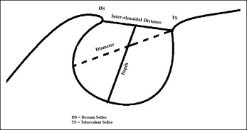
Cephalograms were traced manually on acetate sheets with a 0.5-mm lead pencil in a dark room with conventional methods. Sella turcica was drawn as a U-shaped structure from the tip of the dorsum sellae to that of the tuberculum sellae as seen on the radiograph. The linear dimensions shown in the Figure were measured as follows.
- 1.
Interclinoidal distance: distance from the tip of the dorsum sellae to that of the tuberculum sellae.
- 2.
Depth of sella turcica: distance of a line dropped perpendicular from the line above to the deepest point on the sella floor.
- 3.
Anteroposterior diameter of sella turcica: distance from the tip of the tuberculum sellae to the farthest point on the inner wall of the hypophyseal fossa.

To evaluate and quantify the level of bridging, the standard scoring scale developed by Leonardi et al was used. On the basis of sella dimensions, the bridging was classified into 3 groups.
- 1.
No calcification: this was rated as type I, where the length was either equal to or greater than three fourths of the diameter.
- 2.
Partial calcification: this was rated as type II, where the length was equal to or less than three fourths of the diameter.
- 3.
Complete calcification: this was rated as type III, where only the diaphragm sellae was visible on the radiograph.
To determine the intraexaminer agreement in the identification of the sella turcica bridge, 30 randomly selected lateral cephalometric radiographs were retraced and reevaluated by the principal investigator (B.A.) 2 weeks after the initial analysis. The kappa coefficient value was 0.83, showing a substantial strength of agreement.
SPSS software for Windows (version 19.0; SPSS, Chicago, Ill) was used for the statistical analysis of the data. The chi-square test was performed to test the degree of calcification in both groups. The strength of the association between sella bridging and impacted canines was estimated by calculating the odds ratio. Subjects with partial and complete bridging were grouped in 1 category, and logistic regression analysis was performed. The independent sample t test was used to evaluate differences in the mean sella dimensions between the patients and the controls. P ≤ 0.05 was considered statistically significant.
Results
The mean dimensions of sella turcica in the subjects and the controls are shown in Table I . Independent sample t tests comparing the mean interclinoidal distances between the groups showed a reduced distance among the subjects with impacted canines ( P <0.012). The comparison of mean depths and diameters between the subjects and the controls was insignificant. The patient group was further analyzed for sex dimorphism, which showed no statistically significant difference in sella dimensions ( P >0.05) ( Table II ).
| Sella measurements | Males (n = 11) | Females (n = 20) | P value ∗ |
|---|---|---|---|
| Sagittal interclinoidal distance | 7.22 ± 2.84 | 6.57 ± 1.92 | 0.200 |
| Sella depth | 7.77 ± 1.75 | 8.07 ± 1.36 | 0.120 |
| Sella diameter | 11.40 ± 3.43 | 11.22 ± 2.14 | 0.228 |
Stay updated, free dental videos. Join our Telegram channel

VIDEdental - Online dental courses


