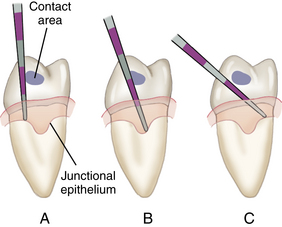
Figure 17-2 A, Incorrect technique for probing the interproximal area. B, Correct technique. C, Incorrect technique.
(Adapted from Perry D, Beemsterboer P, Carranza FA: Techniques and theory of periodontal instrumentation, Philadelphia, 1990, Saunders.)
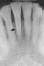
Figure 17-6 Radiograph showing widening of the periodontal ligament associated with occlusal trauma (arrow).
| Class | Description |
|---|---|
| Class I | Beginning involvement. Concavity of furcation can be detected with an explorer or probe, but it cannot be entered. Cannot be detected radiographically. |
| Class II | The clinician can enter the furcation from one aspect with a probe or explorer but cannot penetrate through to the opposite side. |
| Class III | Through-and-through involvement, but the furcation is still covered by soft tissue. A definite radiolucency in the furcation area on a radiograph is visible. |
| Class IV | A through-and-through furcation involvement that is not covered by soft tissue. Clinically it is open and exposed. |
TABLE 17-2 Classification of Mobility
| Class | Description |
|---|---|
| Class I | Tooth can be moved up to 1 mm in any direction. |
| Class II | Tooth can be moved >1 mm in any direction but is not depressible in socket. |
| Class III | Tooth can be moved in a buccolingual direction and is depressible in socket. |
TABLE 17-3 Classification of Fremitus
| Class | Description |
|---|---|
| Class I | Mild vibration or movement detected |
| Class II | Easily palpable vibration but no visible movement |
| Class III | Movement is clearly visible |
Stay updated, free dental videos. Join our Telegram channel
VIDEdental - Online dental courses


