Chapter 7 Panoramic Radiography: Errors Seen in Radiographs
THE NORMAL PANORAMIC RADIOGRAPH
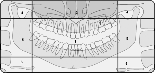
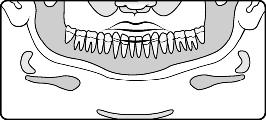
Mind’s Eye View of the Panoramic Radiograph (Diagram 7-1)
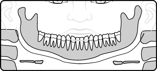
Normal Panoramic Image (Diagram 7-2) Notice that structures have been left out. This is to emphasize that not many structures need to be identified to properly troubleshoot the image for technique errors. In this diagram, the nose and sinus as well as the maxilla are left out because nothing goes wrong here in the perfect image. There is no distinct image of the lips, nose, or ears, nor is there any undesirable air. The teeth, mandible body, ramus, TMJs, and spine are nicely imaged as just described.
STEPS IN TAKING A PANORAMIC RADIOGRAPH
Items left in or on the patient
Improper kVp setting selected:
Bite in the groove in the bite-block.
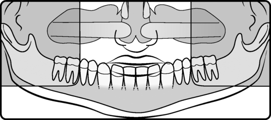
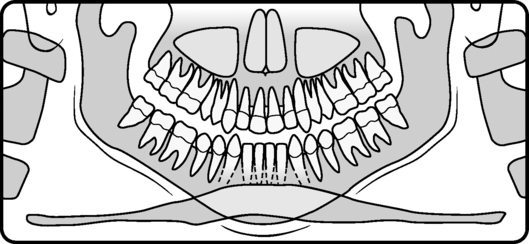
Patient positioned too far forward (Diagram 7-3)
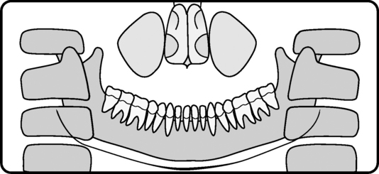
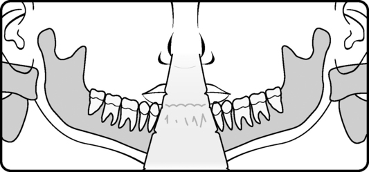
Patient positioned too far back (Diagram 7-4)
Position chin on chin rest, and adjust height of chin rest.
Make sure patient is standing upright with neck and back straight.
Tilt chin downward about 5 degrees.
Check Frankfort plane (ala of nose to tragus of ear) positioning light.
Check canine light to ensure it is still okay.
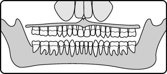
Chin not on chin rest (Diagram 7-5)
Patient slumped or stooped (Diagram 7-6)
Chin tipped too low (Diagram 7-7)
Stay updated, free dental videos. Join our Telegram channel
VIDEdental - Online dental courses


