3 Investigations: Histopathology
Figure 3.1a Pemphigoid.
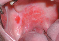
Figure 3.1b Erythroleukoplakia.
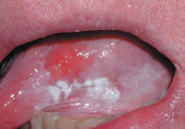
Figure 3.1c White sponge nevus.
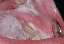
Figure 3.1d White sponge nevus. Typical perinuclear halo 40 x.
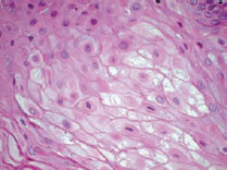
Figure 3.2 Biopsy kit.
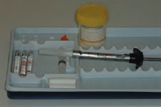
Figure 3.3 Scalpel and punch.
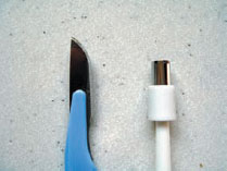
Figure 3.4 Excision biopsy of a lump.
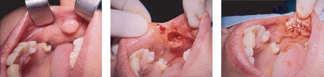
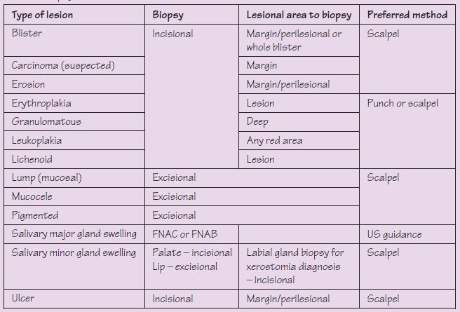
Table 3.1 Biopsy of oral lesions.
Box 3.1 Indications for biopsy
Indications for biopsy include lesions that:
- have neoplastic or potentially malignant features
- are enlarging
- persist > 3 weeks
- are of uncertain etiology
- fail to respond to treatment
- cause concern.
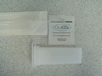
Figure 3.5 Brush biopsy (oral CDx).
Having taken a careful history and completed the clinical examination, the clinician is often in a position to formulate the diagnosis, or at least a list of differential diagnoses. In the la/>
Stay updated, free dental videos. Join our Telegram channel
VIDEdental - Online dental courses


