23: Spread of Dental Infection in the Head and Neck

Chapter 23 Spread of Dental Infection in the Head and Neck

SIGNIFICANT COMPLICATIONS AND IMPLANT DENTISTRY

Few significant complications are associated with implant dentistry. However, identification of potentially life-threatening problems is in the best interest of all concerned. Death in dental practice has been related to air emboli and spread of infection. These complications have a similar foundation: the vascular-lymphatic network and fascial planes. Routes associated with these potential situations are reviewed in this chapter.

The three stages in the development of infection in the head and neck region are the development, extension, and complication stages. The development stage permits the infection to spread through the bone and form an abscess under the periosteum (subperiosteal abscess). This may occur with teeth or implants. When related to an endosteal implant, it is usually associated with a device that is splinted to rigid implants or teeth in the same prosthesis. A subperiosteal implant also may be associated with this complication without the infection coming from within the bone. Most often, before subperiosteal abscess develops, purulent exudate may be found around the permucosal regions of the implant. On rare occasions, a fibrous tissue at the interface between implant and bone may become infected, and the infection may spread through the bone from a retrograde direction. For the most part, implant infection begins from the permucosal region and spreads in an apical direction. Also, an implant placed in an infected immediate extraction site may become involved in retrograde infection.

The extension stage of the development of infection occurs when the subperiosteal abscess penetrates the periosteum and extends to the fascial spaces, producing a cellulitis or fascial abscess. Mandibular subperiosteal implants most often produce this entity in the posterior region, along the posterior body of the mandible. In maxillary subperiosteal or sinus graft surgeries, this stage of infection may develop along the lateral aspect of the maxilla just below the zygomatic arch. The source of the infection is primarily from the maxillary sinus and often has an anaerobic component. On rare occasions, this extension stage has been observed in the submental region from endosteal implants placed through the inferior border of the mandible.

The complication stage results when the infection spreads and causes cavernous sinus thrombosis, brain abscess, neck and mediastinal involvement, pleurisy, or pericarditis. These complications have been reported following sinus surgeries, primarily when the antrum was infected at the time of surgery.

The principal routes for the spread of dental infection are through the following four mechanisms:

Subperiosteal implants often extend beyond muscle attachments or fascial planes. Infections associated with primary struts should be closely supervised and aggressively treated because the infection may spread by continuing beyond these structures. Endosteal implants are most often positioned beyond the apices of natural teeth. As a result, infections beyond their apices may perforate bone beyond the usual limiting borders associated with the roots of teeth and may result in subcutaneous rather than submucosal infection.

PATHOLOGIC ENTITIES

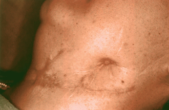
The definitions of specific terms that describe infections of the head and neck provide keys to methodology of treatment and improved communications. An acute cellulitis involves diffuse inflammation of the areolar connective tissue and loose subcutaneous tissues. Lymphadenitis is a condition in which the regional lymph nodes become inflamed, enlarged, and tender. The node may become suppurated, break through the capsule, and involve the surrounding tissues. Abscess formation results when tissues break down and leukocytes die, thus forming pus. Staphylococci and streptococci are the usual bacteria involved in this process; however, a more varied population is prevalent in sinus infections. Phlegmon is any cellulitis that does not go on to suppuration. In this condition, the inflammatory infiltration of the subcutaneous tissue leads to accumulation of foul-smelling, brownish exudate. Hemolytic streptococci are usually present. A chronic cellulitis follows an acute cellulitis and may be the result of inadequate treatment or a subvirulent organism with no suppuration. A chronic abscess is a well-encapsulated entity caused by a subvirulent organism. In this case, a bone sequestrum or retained root tip is the more common source of the infection. A chronic skin fistula is a sign of retained focus of infection and, in some cases, a more serious condition of bone and bone marrow inflammation called osteomyelitis (Figure 23-1). This has been observed in the mandible and is associated with infection of both endosteal and subperiosteal implants in patients who have poor dental awareness and lack of implant maintenance. A noma starts as a gangrenous stomatitis and spreads to adjacent bone and muscles, causing lysis and necrosis of tissue. This rare condition perforates the cheek, floor of the mouth, or both, and is usually seen in debilitated individuals.

MAXILLARY INFECTIONS

Acute suppurative periapical abscesses in the maxilla may penetrate the alveolar bone and form a subperiosteal abscess under the periosteum. If the abscess penetrates the periosteum above the attachment of the buccinator muscle, it causes a buccal abscess.

If the abscess continues to spread, it may involve the skin, forming a cutaneous abscess. The infection may progress superiorly and involve the temporal region. Downward extension may involve the submandibular space. If the abscess perforates the buccal alveolar bone and periosteum below the attachment of the buccinator muscle, the abscess wall appears intraorally in the buccal vestibule and forms a gingival or alveolar abscess.

Palatal extension of the infection is rare. Usually it is from lingual roots of the upper molars, forming a palatal abscess. On occasion, an implant may perforate the palatal bone and cause an infection that may be evident at a later date.

The maxillary sinus may become invaded by an infection of the maxillary teeth, resulting in an acute sinusitis. Radiographs reveal a clouded sinus resulting from accumulation of inflammatory exudates within the sinus cavity. Sinus graft surgeries may penetrate an already infected antrum, or one that becomes infected shortly after surgery, with subsequent extension of the infection into the whole implanted region. The risk of sinus infection immediately following surgery is approximately 3%.

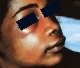
The loose, fat-containing connective tissue of the lips and cheeks is continuous and is traversed by the muscles of facial expression, which arise from the bones of the face, traverse the subcutaneous tissue, and end in the skin. These muscles, with their thin perimysium, play a role in directing the spread of infection. Dental or implant abscesses that erode and perforate the facial alveolar compact bone sometimes find their way through the subcutaneous tissue and produce remarkable swelling of the upper lip and cheek and may spread to the lower and upper eyelids due to the lack of fascial barriers on the face (Figure 23-2).

Most often, maxillary dental infections involve four regions of the maxilla: the upper lip, canine fossa, buccal space, and infratemporal space. The most common maxillary implant-associated infection is in the buccal space (Figure 23-3).

image

Figure 23-3 Buccal surgical space infection with spread to the submandibular space.

(From Hupp JR, Tucker MR, Ellis E: Contemporary oral and maxillofacial surgery, ed 5, St Louis, 2009, Mosby.)

Stay updated, free dental videos. Join our Telegram channel

Jan 7, 2015 | Posted by in Implantology | Comments Off on 23: Spread of Dental Infection in the Head and Neck

VIDEdental - Online dental courses

Get VIDEdental app for watching clinical videos