2
The basics: the peri-implant mucosa
After implant placement, a delicate mucosal attachment is established. The peri-implant mucosa is sealed to the implant surface to protect the bone tissue, and to prevent the penetration of micro-organisms and their products. Limited data exist in humans. Most of the following information is extrapolated from animal studies. Thus, data on healing time might not always be directly transferable to the clinical situation.
The peri-implant mucosa results from the healing process of the soft tissues surrounding the implant following the closure of the flap around the transgingival part of the implant.
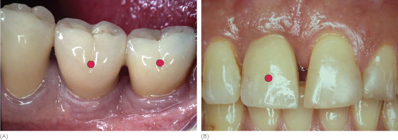
From a clinical point of view, the outer surface of the peri-implant mucosa is covered by a keratinized oral epithelium. It has a pink color and a firm consistency, and does not differ from the clinical appearance of the gingiva (Fig. 2.1A, B). The peri-implant mucosa clinical dimension tends to be thicker and lower in height than the gingiva surrounding teeth.
Figure 2.1 (A,B) Clinical appearance of the peri-implant mucosa. Red circles indicate the implant-supported prosthesis.
From a histological point of view, compared to the periodontal model, the dental implant model has the following main features (Fig. 2.2):
- lack of cementum
- lack of periodontal ligament
Stay updated, free dental videos. Join our Telegram channel
VIDEdental - Online dental courses


