15
Oral pathology and oral surgery
J.G. Meechan
Chapter contents
15.2 Lesions of the oral soft tissues
15.2.3 Vesiculobullous lesions
15.3.2 Developmental conditions
15.4 Oral manifestations of systemic disease
15.5.3 Surgical aids to orthodontics
15.5.5 Cysts interfering with eruption
15.5.6 Treatment of acute orofacial infection
15.5.7 Autotransplantation of teeth
15.7.5 Excision biopsy of non-attached mucosa
15.7.6 Excision biopsy of attached gingiva/palate
15.1 Introduction
The incidence of pathological conditions of the mouth and perioral structures varies between children and adults. For example, mucocoeles are more common in the young, whereas squamous cell carcinomas occur more frequently in older individuals. The management of pathology in the child differs from that in the adult. Growth and development may be affected by the disease or by its treatment. On a more practical basis, anaesthetic considerations for surgical treatment of simple pathological conditions can make management more complex in the young patient. This chapter deals with those conditions that occur exclusively or more commonly in children. It is not an exhaustive guide to paediatric oral pathology, for which readers should refer to oral pathology textbooks. Surgical treatment of the simpler conditions is discussed in the oral surgery section of this chapter (Section 15.5).
15.2 Lesions of the oral soft tissues
Conditions affecting the oral mucosa and associated soft tissues can be classified as follows: infections, ulcers, vesiculobullous lesions, white lesions, cysts, and tumours.
15.2.1 Infections
Viruses, bacteria, fungi, or protozoa may cause infections of the oral mucosa. Odontogenic infections will be discussed in Section 15.5.6.
Viral infections
Herpetic infections
Primary herpes simplex infection This condition usually occurs in children between the ages of 6 months and 5 years. Circulating maternal antibodies usually protect young babies. The symptoms, signs, and treatment are covered in Chapter 11.
Secondary herpes simplex infection Secondary infection with herpes simplex usually occurs at the labial mucocutaneous junction and presents as a vesicular lesion that ruptures and produces crusting (see Chapter 11).
Herpes varicella-zoster Shingles, which is caused by the varicellazoster virus, is much more common in adults than in children. The vesicular lesion develops within the peripheral distribution of a branch of the trigeminal nerve.
Chickenpox, a more common presentation of varicella-zoster in children, produces a vesicular rash on the skin. The intra-oral lesions of chickenpox resemble those of primary herpetic infection. The condition is highly contagious.
Mumps
Mumps produces a painful enlargement of the parotid glands. It is usually bilateral. The causative agent is a myxovirus. Associated complaints include headache, vomiting, and fever. Symptoms last for about a week and the condition is contagious.
Measles
The intra-oral manifestation of measles occurs on the buccal mucosa. The lesions appear as white speckling surrounded by a red margin and are known as Koplick’s spots. The oral signs usually precede the skin lesions and disappear early in the course of the disease. The skin rash of measles normally appears as a red maculopapular lesion. Fever is present and the disease is contagious.
Rubella
Rubella (German measles) does not usually produce signs in the oral mucosa; however, the tonsils may be affected. Protection against the diseases of mumps, measles, and rubella can be achieved by vaccinating children with MMR vaccine in their early years.
Herpangina
This is a Coxsackie virus A infection. It can be differentiated from primary herpetic infection by the different location of the vesicles, which are found in the tonsillar or pharyngeal region. Herpangina lesions do not coalesce to form large areas of ulceration. The condition is short-lived.
Hand, foot, and mouth disease
This Coxsackie virus A infection produces a maculopapular rash on the hands and feet. The intra-oral vesicles rupture to produce painful ulceration. The condition lasts for 10–14 days.
Infectious mononucleosis
The Epstein–Barr virus causes this condition. It is not uncommon amongst teenagers. The usual form of transmission is by kissing. Oral ulceration and petechial haemorrhage at the hard–soft palate junction may occur. There is lymph node enlargement and associated fever. There is no specific treatment. It should be noted that prescription of ampicillin and amoxicillin (amoxycillin) can cause a rash in those suffering from infectious mononucleosis. These antibiotics should be avoided during the course of the disease. Treatment of the above viral illnesses is symptomatic and relies on analgesia and maintenance of fluid intake. It must be remembered that aspirin should be avoided in children under 12 years of age (see later).
Human papillomavirus
This is associated with a number of tumour-like lesions of the oral mucosa, which are discussed in Section 15.2.6.
Bacterial infections
Staphylococcal infections
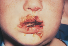
Staphylococci and streptococci may cause impetigo. This can affect the angles of the mouth and the lips (Fig. 15.1). It presents as crusting vesiculobullous lesions. The vesicles coalesce to produce ulceration over a wide area. Pigmentation may occur during healing. The condition is self-limiting, although antibiotics may be prescribed in some cases. Staphylococcal organisms can cause osteomyelitis of the jaws in children. Although the introduction of antibiotics has reduced the incidence of severe forms of the condition, it can still be devastating. In addition to aggressive antibiotic therapy, surgical intervention is required to remove bony sequestrae.
Streptococcal infection
Streptococcal infections in childhood vary from a mucopurulent nasal discharge to tonsillitis, pharyngitis, and gingivitis. Scarlet fever is a β–haemolytic streptococcal infection consisting of a skin rash with maculopapular lesions of the oral mucosa. It is associated with tonsillitis and pharyngitis. The tongue shows characteristic changes from a strawberry appearance in the early stages to a raspberry-like form in the later stages.
Congenital syphilis
Congenital syphilis is transmitted from an infected mother to the fetus. Oral mucosal changes such as rhagades, which is a pattern of scarring at the angle of the mouth, may occur. In addition, this disease may cause characteristic dental changes in the permanent dentition. These include Hutchinson’s incisors (the teeth taper towards the incisal edge rather than the cervical margin) and mulberry molars (globular masses of enamel over the occlusal surface).
Figure 15.1 Bacterial infection on the lips of an immunocompromised child. (Reproduced from Dental Update (ISSN 0305-5000), by permission of George Warman Publications (UK) Ltd.)
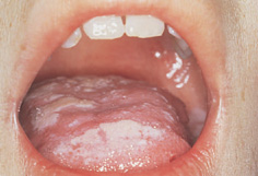
Figure 15.2 Oral candidiasis in an immunocompromised child undergoing chemotherapy for acute lymphoblastic leukaemia. (Reproduced from Dental Update (ISSN 0305-5000), by permission of George Warman Publications (UK) Ltd.)
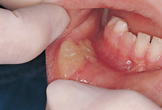
Figure 15.3 Ulceration of the lower lip produced by biting while still anaesthetized from an inferior block.
Tuberculosis
Tuberculous lesions of the oral cavity are rare. However, tuberculous lymphadenitis affecting submandibular and cervical lymph nodes is occasionally seen. These present as tender enlarged nodes, which may progress to abscess formation with discharge through the skin. Surgical removal of infected glands produces a much neater scar than that caused by spontaneous rupture through the skin if the disease is allowed to progress.
Cat-scratch disease
This is a self-limiting disease which presents as an enlargement of regional lymph nodes. The nodes are painful and enlargement occurs up to 3 weeks following a cat scratch. The nodes become suppurative and may perforate the skin. Treatment often involves incision and drainage.
Fungal infections
Candida
Neonatal acute candidiasis (thrush) contracted during birth is not uncommon. Young children may develop the condition when resistance is lowered or after antibiotic therapy (Fig. 15.2). Easily removed white patches on an erythematous or bleeding base are found. Treatment with nystatin or miconazole is effective (those under 2 years of age should receive 2.5mL of a miconazole gel (24mg/mL) twice daily; 5mL twice daily is prescribed for those under 6 years of age, and 5mL four times daily for those over 6 years of age).
Actinomycosis
Actinomycosis can occur in children. It may follow intra-oral trauma including dental extractions. The organisms spread through the tissues and can cause dysphagia if the submandibular region is involved. Abscesses may rupture onto the skin and long-term antibiotic therapy is required. Penicillin should be prescribed and maintained for at least 2 weeks following clinical cure.
Protozoal infections
Infection by Toxoplasma gondii may occasionally occur in children, with the principal reservoir of infection being cats. Glandular toxoplasmosis is similar in presentation to infectious mononucleosis and is found mainly in children and young adults. There may be a granulomatous reaction in the oral mucosa and there can be parotid gland enlargement. The disease is self-limiting, although an antiprotozoal such as pyrimethamine may be used in cases of severe infection.
15.2.2 Ulcers
Traumatic ulceration of the tongue, lips, and cheek may occur in children, especially after local anaesthesia has been administered (Fig. 15.3).
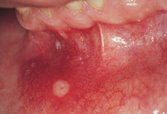
Recurrent aphthous oral ulceration not associated with systemic disease is often found in children (Fig. 15.4). One or more small ulcers in the non-attached gingiva may occur at frequent intervals. In the young child the symptoms may be mistaken for toothache by a parent. The majority of aphthous ulcers in children are of the minor variety (less than 5mm in diameter). These usually heal within 10–14 days. Treatment other than reassurance is often unnecessary; however, topical steroids may be prescribed in severe cases. Older children may benefit from the use of antiseptic rinses to prevent secondary infection. In the absence of a history of major aphthous ulceration any ulcer lasting for longer than 2 weeks should be regarded with suspicion and biopsied.
Figure 15.4 Minor aphthous ulceration. (Reproduced by kind permission of Informa Healthcare.)
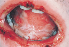
Figure 15.5 Erythema multiforme in a teenager.
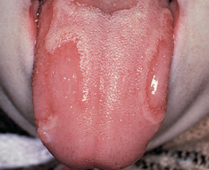
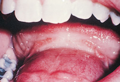
Figure 15.6 Geographic tongue. (Reproduced by kind permission of Informa Healthcare.)
15.2.3 Vesiculobullous lesions
Vesiculobullous lesions cause ulcers in the later stages of such conditions. Viral causes have been mentioned above. Similarly, conditions such as epidermolysis bullosa and erythema multiforme can produce oral ulceration in children. The major vesiculobullous conditions, such as pemphigus and pemphigoid, are rare in young patients. Epidermolysis bullosa is a term that covers a number of syndromes, some of which are incompatible with life. The skin is extremely fragile and mucosal involvement may occur. The act of suckling may induce bullae formation in babies. In older children effective oral hygiene may be difficult as even mild trauma can produce painful lesions.
The oral lesions of erythema multiforme usually affect the lips and anterior oral mucosa (Fig. 15.5). There is initial erythema followed by bullae formation and ulceration. The pathogenesis of the condition is still unclear; however, precipitating factors include drug therapy and infection. Treatment involves the use of steroids and oral antiseptic and analgesic rinses to ease the pain.
15.2.4 White lesions
Trauma of either a chemical or physical nature (e.g. burns and occlusal trauma) can cause white patches intra-orally.
White spongy naevus
White spongy naevus (also known as oral epithelial naevus) is a rough folded lesion that can affect any part of the oral mucosa. It often appears in infancy. It is benign.
Leucoedema
This is a folded white translucent appearance found in children of races who exhibit pigmentation of the oral mucosa. It is considered a variation of normal.
Candidiasis
The white patches of acute fungal candidiasis mentioned above are readily removed, in contrast to the white lesions discussed here.
Geographic tongue
This condition may be seen in children. It is normally symptomless, although some patients complain of discomfort with spicy foods. Areas of the tongue appear shiny and red as a result of loss of filiform papillae (Fig. 15.6). These red patches are surrounded by white margins. These areas disappear before reappearing in other regions of the tongue. The condition is benign and requires no treatment apart from reassurance to the child and parent.
15.2.5 Cysts
Mucocoeles
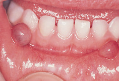
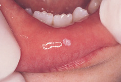
The peak incidence of mucocoeles is in the second decade of life; however, they are not uncommon in younger children (Fig. 15.7) including neonates. Mucocoeles are caused by trauma to minor salivary glands or ducts and are often located on the lower lip. They are the most common non-infective cause of salivary gland swelling in children. Salivary tumours are rare in this age group.
Ranula
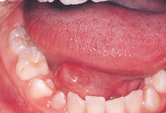
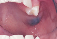
This appears a bluish swelling of the floor of the mouth (Fig. 15.8). It is essentially a large mucocoele. It may arise from part of the sublingual salivary gland.
Figure 15.7 Bilateral mucocoeles in a 3-year-old girl. (By kind permission of the Journal of Dentistry for Children.)
Figure 15.8 A ranula in a 14-year-old girl.
Figure 15.9 A dermoid cyst.
Figure 15.10 Squamous cell papilloma in a 9-year-old girl. (Reproduced from Dental Update (ISSN 0305-5000) by permission of George Warman Publications (UK) Ltd.)
Bohn’s nodules
These gingival cysts arise from remnants of the dental lamina. They are found in neonates and usually disappear spontaneously in the early months of life.
Epstein’s pearls
These small cystic lesions are located along the palatal midline. They are thought to arise from trapped epithelium in the palatal raphe. They are present in about 80% of neonates and disappear within a few weeks of birth.
Dermoid cysts
These are rare lesions of the floor of the mouth. They appear as intra-oral and submental swellings (Fig. 15.9). They are derived from epithelial remnants remaining after fusion of the mandibular processes.
Lymphoepithelial cyst
In the past this was termed branchial arch cyst as it was thought to arise from epithelial remnants of a branchial arch. Lymphoepithelial cysts are normally found in the sternomastoid region, although they can present in the floor of the mouth. Histologically, the cyst wall contains lymph tissue. The tissue of origin is thought to be salivary epithelium.
Thyroglossal cyst
This cyst, which arises from the thyroglossal duct epithelium, may present intra-orally. However, the mouth is a rare site. Most arise in the region of the hyoid bone.
15.2.6 Tumours
Congenital epulis
This is a rare lesion that occurs in neonates. It normally presents in the anterior maxilla. It consists of granular cells covered by epithelium and is thought to be reactive in nature. This is a benign lesion and simple excision is curative.
Melanotic neuroectodermal tumour
This rare tumour occurs in the early months of life, usually in the maxilla. The lesion consists of epithelial cells containing melanin with a fibrous stroma. Some localized bone expansion may occur. The condition is benign and simple excision is curative.
Squamous cell papilloma
This is a benign condition which occurs in children. The small pedunculated cauliflower-like growths, which vary in colour from pink to white (Fig. 15.10), are usually solitary lesions. They are caused by the human papillomavirus.
Verruca vulgaris
This condition, also known as the common wart, may present as solitary or multiple intra-oral lesions. These may be associated with skin warts. They are caused by the human papillomavirus.
This is a rare condition also known as Heck’s disease. It is associated with human papillomavirus. It presents as multiple small elevations of the oral mucosa, especially in the lower lip.
Fibroepithelial polyp
This is a fairly common lesion that presents as a firm pink lump. It normally affects the buccal mucosa at the occlusal level. These lesions are caused by trauma. They are usually symptomless unless further traumatized and are easily removed.
Fibrous epulis
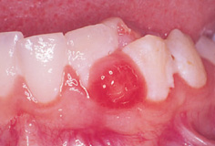
This presents as a mass on the gingiva. The colour varies from pink to red depending on the degree of vascularity of the lesion (Fig. 15.11). It consists of an inflammatory cell infiltrate and mature fibrous tissue; occasionally a calcified variant is found. Surgical excision is curative.
Pyogenic granuloma
These commonly occur on the gingiva, usually in the anterior maxilla. They are probably a reaction to chronic trauma, especially from subgingival calculus. As a result of their aetiology they have a tendency to recur after removal.
Peripheral giant cell granuloma
This dark-red swelling of the gingiva can occur in children. It often arises interdentally. Radiographs may reveal some loss of the interdental crest. The central giant cell granuloma (see Section 15.3.4) shows much greater bone destruction. This condition is thought to be a reactive hyperplasia. Unless excision is complete the granuloma will recur.
Haemangiomas
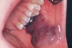
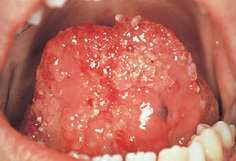
Haemangiomas are relatively common in children. They are malformations of blood vessels. They are divided into cavernous and capillary variants, although some lesions contain elements of both. Capillary haemangiomas may present as facial birthmarks. The cavernous haemangioma is a hazard during surgery if involved within the surgical site. It is a large blood-filled sinus that will bleed profusely if damaged (Fig. 15.12). The extent of a cavernous haemangioma can be established prior to surgery using either angiography or MRI scanning. Small haemangiomas are readily treated by excision or cryotherapy. Larger lesions are amenable to laser therapy.
Sturge–Weber syndrome
Sturge–Weber angiomatosis is a syndrome consisting of a haemangioma of the leptomeninges with an epithelial facial haemangioma closely related to the distribution of branches of the trigeminal nerve. Mental deficiency, hemiplegia, and occular defects can occur. Intra-oral involvement may interfere with the timing of eruption of the teeth (both early and delayed eruption have been reported).
Lymphangiomas
Lymphangiomas are benign tumours of the lymphatics. The vast majority are found in children. The head and neck region is a common site (Fig. 15.13). The cystic hygroma is a variant that appears as a large neck swelling which may extend intra-orally to involve the floor of the mouth and tongue.
Neurofibromas
These may present as solitary or multiple lesions. They are considered to be hamartomas (a haphazard arrangement of tissue). They present intra-orally as mucosal swellings on the tongue or gingivae. Multiple oral neuromas are a feature of multiple endocrine neoplasia syndrome. As the oral signs may precede the development of more serious aspects of this condition (such as carcinoma of the thyroid), children presenting with multiple lesions should be referred to an endocrinologist.
Figure 15.11 A fibrous epulis in a 10-year-old girl (a pyogenic granuloma appears similar).
Figure 15.12 Cavernous haemangioma of the buccal mucosa. (Reproduced by kind permission of Informa Healthcare.)
Figure 15.13 Lymphangioma of the tongue and floor of the mouth. (By kind permission of Professor C. Scully.)
Orofacial granulomatosis (OFG)
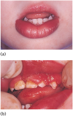
OFG is not a tumour in the true sense, nor a distinct disease entity, but describes a clinical appearance. Typically, there is diffuse swelling of one or both lips and cheeks, cobble-stoning and tags of the buccal reflected mucosa, full width gingivitis, and oral ulceration (Fig. 15.14). This may represent a localized disturbance as a manifestation of an allergic reaction to foodstuffs, toothpaste, or even dental materials. Alternatively, the appearance may be due to an underlying systemic condition such as sarcoidosis or Crohn’s disease.
Melkersson–Rossenthal syndrome
This condition generally begins during childhood. It consists of chronic facial swelling (usually the lips), facial nerve paralysis, and a fissured (scrotal) tongue.
Malignant tumours of the oral soft tissues
Epithelial tumours
Malignant tumours of the oral epithelium, such as squamous cell carcinoma, are rare in children. Malignant salivary neoplasms are also uncommon, although muco-epidermoid carcinomas have been reported in young patients.
Lymphomas
Hodgkin’s and non-Hodgkin’s lymphomas can occur in children, but they are relatively rare in the paediatric age group. An exception is Burkitt’s lymphoma, which is endemic in parts of Africa and affects those under 14 years of age. Indeed, in these areas the condition accounts for almost half of all malignancies in children. Burkitt’s lymphoma is multifocal, but a jaw tumour (more often in the maxilla) is often the presenting symptom. Burkitt’s lymphoma is strongly linked to the Epstein–Barr virus as a causal agent.
Figure 15.14 (a) Swelling of the lower lip and (b) attached mucosa and gingiva in a 3-year-old girl with orofacial granulomatosis.
Rhabdomyosarcomas
These malignant tumours of skeletal muscle present in patients around 9–12 years of age. The usual site is the tongue. Metastases are common and the prognosis is poor.
15.3 Lesions of the jaws
These can be divided into cysts, developmental conditions, osteodys-trophies, and tumours.
15.3.1 Cysts
Eruption
Eruption cysts are really dentigerous cysts, which present as swellings of the alveolar mucosa. They may precede the eruption of both primary and permanent teeth (Fig. 15.15). When filled with blood they are often called eruption haematomas. The treatment of eruption cysts is discussed in Section 15.5.5.
Dentigerous
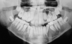
This is the most common jaw cyst in children (Fig. 15.16). Its origin is the reduced enamel epithelium, and attachment to the tooth occurs at the amelocemental junction. There are often no symptoms, but eruption of the affected tooth will be prevented. The treatment of dentigerous cysts is discussed in Section 15.5.5.
Radicular
These cysts, which are related to the apex of a non-vital tooth, do occur in children although they are rare in the primary dentition. They are often symptomless and are discovered radiographically. Extraction, apicectomy, or conventional endodontics will effect a cure. Lateral periodontal cysts are very rare in children.
Keratinizing odontogenic tumour
This lesion, which was formerly known as the odontogenic kerato-cyst, is the most aggressive of the jaw cysts. Rates of recurrence of around 60% have been reported because fragments remaining after subtotal removal will regenerate. These cysts may be found in children and may be associated with the basal cell naevus (Gorlin–Goltz) syndrome. The odontogenic tumours associated with this syndrome appear in the first decade of life, whereas the syndromic basal cell carcinomas are rare before puberty. Other signs and symptoms include multiple basal cell carcinomas, bifid ribs, calcification of the falx cerebri, hypertelorism, and frontal and temporal bossing.
Figure 15.15 Eruption cyst prior to the appearance of the upper permanent molar.
Figure 15.16 Radiographic appearance of a dentigerous cyst associated with a lower second premolar. (Reproduced from Dental Update (ISSN 0305-5000), by permission of George Warman Publications (UK) Ltd.)
