Radiographic Assessment and Interpretation
MULTIPLE CHOICE QUESTIONS WITH A RADIOGRAPHIC IMAGE
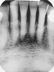
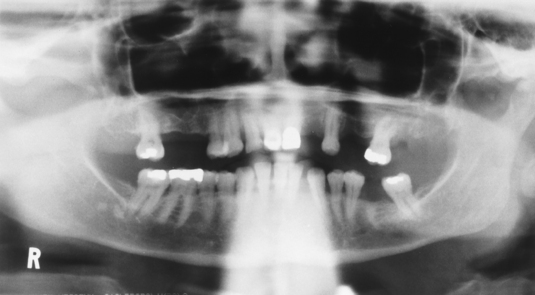
302. This patient is a 60-year-old man with markedly shortened crowns. He does not work in an environment where particulate matter or acid-containing fumes can pollute the air. He has no known eating disorders and is healthy systemically. By what process have the crowns acquired this appearance?
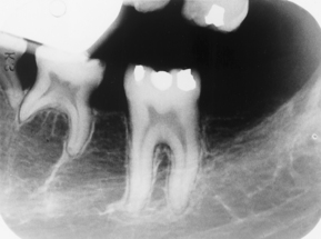
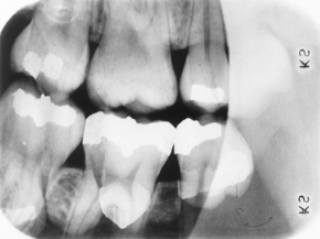
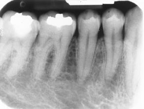
303. Observe the bifurcation area of these three molars. All have the same round, radiopaque, anomalous appearance. Note the overlap of the contacts. What term best describes this?
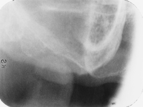
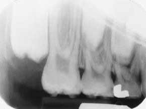
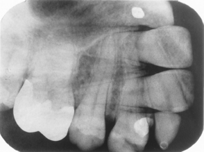
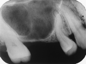
305. At least two errors are in this edentulous maxillary posterior periapical view. Select the best choice.
308. This patient is a 32-year-old white woman. This was the only lesion she had, and the adjacent teeth were vital. The condition we see here is:
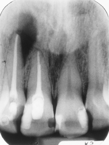
309. This patient first had the endo done after a long period of abscess and fistula formation. Then the lateral incisor reabscessed and an apicoectomy and curettage were done. Currently the patient is asymptomatic and clinically there is a scar but the area appears well healed. What is your assessment of the periapical radiolucent area at the apex of the lateral incisor?
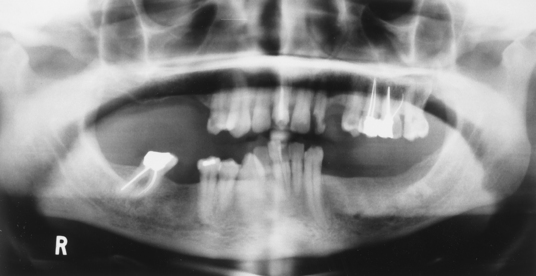
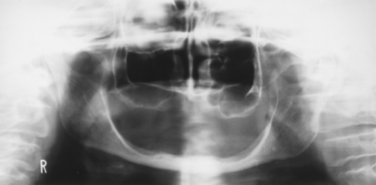
311. In this edentulous patient we can see a number of errors. The most complete and accurate list is:
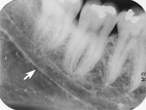
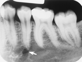
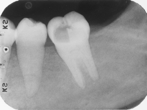
313. The 2nd premolar is vital and asymptomatic, and the patient is a black female. Identify the radiolucency to which the arrow is pointing.
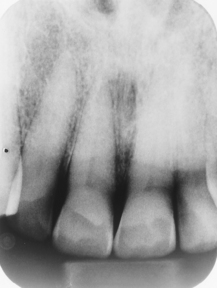
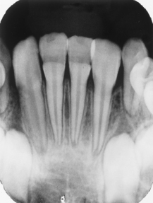
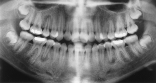
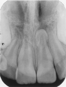
315. We are interested in the central incisors of this 7-year-old boy who has had a lot of fevers during a certain period of his life. What condition affects the central incisors, and when during his life did this occur?
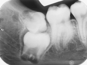
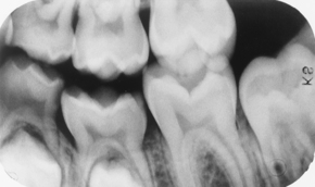
316. Here we see a very good radiograph of the 3rd molar region. List the anomalies seen in this radiograph.
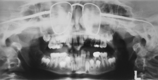
317. Notice that there are at least two, possibly three, missing permanent teeth with the retention of at least one or two primary teeth. Among the following list, what is the most likely diagnosis?
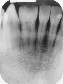
318. This patient is a 72-year-old man. Notice that the pulp and root canal spaces are significantly diminished. What is the cause of this?
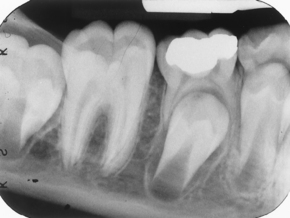
320. This young adult is missing her 1st premolars; there is also a technique error in this film. Which choice best represents this case?
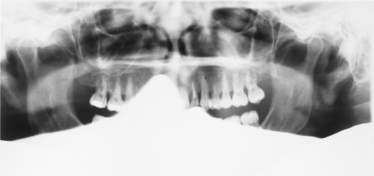
324. Note that the lips are slightly open and the tongue is not quite up against the palate; the lead apron may have ridden up very slightly on the shoulder. Several additional errors are in this panoramic radiograph. See if you can find them all.
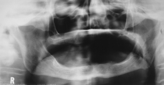
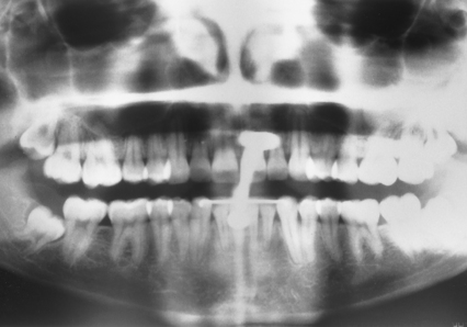
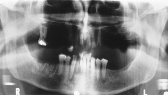
325. This patient’s lips are closed (you can see them), and the tongue is against the palate. There are, however, several errors. What are they?
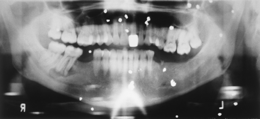
326. This patient survived a little run-in he had with farmer Brown’s shotgun. This is a bit tricky: Identify the metallic objects that have produced (a) ghost image(s).
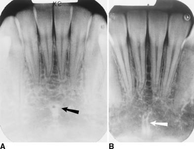
328. In part A, there is a black arrow and in part B a white arrow. Together they depict what anatomic structures?
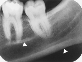
329. In this periapical radiograph there are two white arrowheads. To what structures do they point?
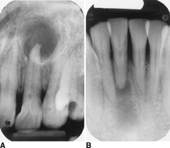
330. The maxillary lateral incisor (part A) and the mandibular central incisor (part B) both have periapical radiolucencies in this 59-year-old white man. Read the following question carefully: Select the best choice stating the nature of the periapical lesion and the one tooth with the visible cause identified.
A. Periapical lesion of pulpal origin (abscess, cyst, granuloma) both teeth; trauma to lower central incisor
B. Periapical lesion of pulpal origin (abscess, cyst, granuloma) both teeth; dens in dente maxillary lateral incisor
331. Regarding this image, select the one most accurate choice listing what can be seen in this image.
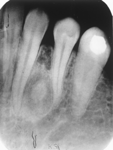
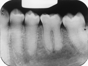
332. We are considering the radiolucent lesion between the lower premolars. Based on this radiograph, what would be your most likely clinical diagnosis before biopsy?
333. This patient is a 26-year-old woman with kidney disease. Note the ground-glass pattern of the alveolar bone and loss of the lamina dura. Select the most likely diagnosis.
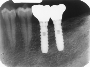
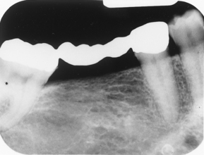
335. Observe the radiograph of this fixed 4 unit prosthesis (bridge). What materials is the prosthesis made of?
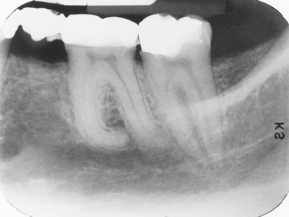
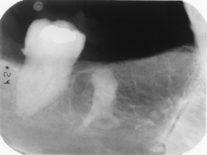
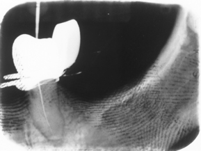
336. This patient has a history of a fractured mandible. What do you make of what we see at the apex of the 2nd (most posterior) molar?
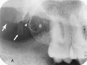
338. First let’s get oriented. Note the sigmoid notch and coronoid process of the mandible (black A) and the maxillary tuberosity (white B). Select the correct combination of answers listed from the most posterior (large arrow), the middle (small arrow), and the most anterior (arrowhead).
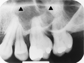
339. Note the many accessory canals in the lateral walls (anterolateral and posterolateral) of the maxillary sinus and the malar process of the maxilla superimposed on the 2nd molar. This question deals with only the structure indicated by the arrowheads. Select the best choice.
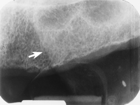
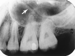
340. Look closely at the maxillary sinus. The arrow points to a radiolucent line bound by a more radiopaque line on each side that cuts diagonally across the maxillary sinus. This is:
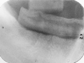
341. Because of the blackness of the fingerprints, what chemical do you think contaminated this film?
342. For this radiograph, match the descriptive term that indicates the problem; after that, list the cause.
344. Oh yes, you can believe it! Stuff like this happens. Okay, select the most complete list of errors.
346. Read this one carefully. The patient is a middle-aged black woman. Match the radiographic findings with the associated disorder.
347. This patient had a tonsillectomy years ago and has the following symptoms: a sensation like a fishbone stuck in the throat on swallowing and occasional lightheadedness when turning the head. The condition depicted here is caused by:
348. This patient is a 47-year-old black woman. There were some periapical radiopacities in the anterior region, which is obscured in this case. The diagnosis is:
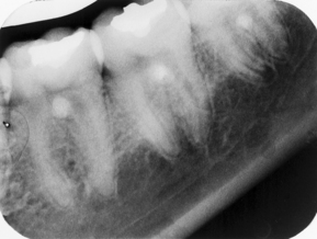
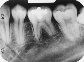
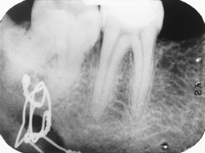
349. Amazingly, three of the four 2nd premolars were non-vital in this 14-year-old Asian teenager. All four 2nd premolars were affected by the same clinical finding. What could this be?
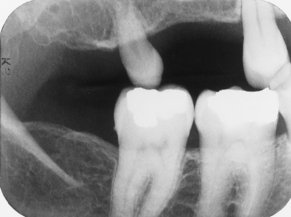
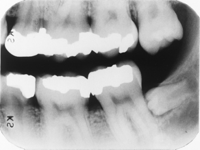
352. Note the extruded maxillary 3rd molar. What term(s) best describe(s) the most distal mandibular tooth? Note that the 1st and 2nd molars are present and no teeth have been extracted.
Stay updated, free dental videos. Join our Telegram channel

VIDEdental - Online dental courses


