13 Pigmented lesions
Figure 13.1a Causes of pigmentation.
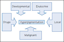
Figure 13.1b Pigmentation diagnosis.
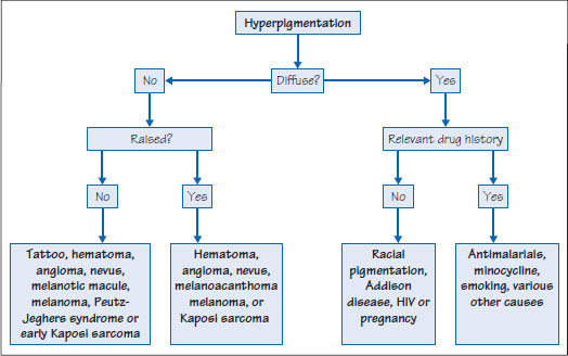
Figure 13.2 Bismuth induced black tongue.
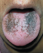
Figure 13.3a Hairy tongue. Note central discoloration.
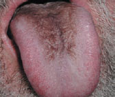
Figure 13.3b Hairy tongue.
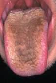
Table 13.1 Causes of isolated hyperpigmented lesions.
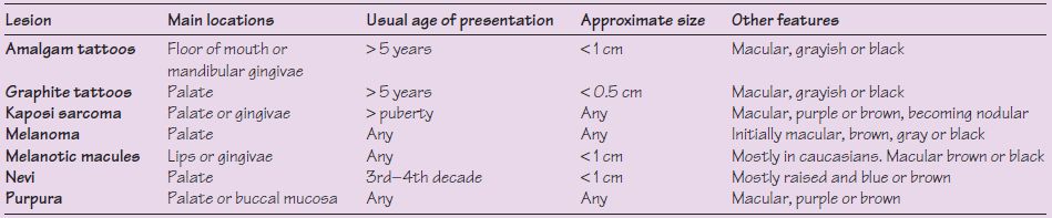
The oral mucosa is usually not very pigmented despite the fact that it has the same melanocyte density as skin. Pigmentation may be superficial (extrinsic) or intrinsic, isolated lesions or generalized, and ranges from light brown to blue-black, gray, red, or purple – the color depending on the pigment and its depth. Melanin pigment is brown, but can impart a black, brown, blue, or green color to the eye. Vascular lesions tend to be red, purple or blue, but hemorrhage can lead to red, purple, brown or other colors (Tables 13.1 and 13.2).
Intrinsic hyperpigmentation can have a range of causes (Figures 13.1a and b).
Superficial discoloration
Superficial brown discoloration of tongue and teeth, which is easily removed and of little consequence is commonly caused by habits (e.g. cigarette smoking, tobacco or betel chewing); beverages (e.g. coffee, tea and red wine); foods (e.g. beet, liquorice); or by drugs (e.g. iron, chlorhexidine, bismuth) (Figure 13.2). Hairy tongue, often referred to as black hairy tongue, may also appear brown, white, green, or pink.
Table 13.2 Causes of oral pigmentation.
| Causes | Comments |
Stay updated, free dental videos. Join our Telegram channel
VIDEdental - Online dental courses


