Chapter 12
Endodontic Surgery
Periradicular surgical procedures (PRS) are used to treat diseases and conditions of the tooth root that cannot be treated with orthograde root canal treatment. The majority of these surgical procedures will involve resection of the root apex (apicectomy) and retrograde obturation of the root canal to address persistent disease that has not resolved following an acceptable root canal treatment.
Indications for Periapical Surgery
An apicectomy is indicated when conventional non-surgical root canal treatment with orthograde root canal filling has failed or cannot be performed, and the tooth is associated with clinical symptoms or signs of continuing periradicular disease. A surgical approach cannot substitute non-surgical orthograde root canal treatment and should not be considered as the primary option to treat teeth with associated apical pathology, but seen as a supplement to endodontic treatment. Indications and contraindications for undertaking PRS are outlined in Table 12.1.
Table 12.1 Indications and contraindications for surgical endodontics. (Adapted from ESE guidelines 2006.)
| Indications for surgical endodontics | Contraindications for surgical endodontics |
|---|---|
| Radiological findings of periradicular periodontitis and/or symptoms associated with a canal where an obstruction cannot be removed or will result in damage Extruded material with clinical or radiological findings of apical periodontitis and/or symptoms over a prolonged period Persisting or emerging disease following root canal treatment where retreatment is inappropriate Perforation of the root or pulp chamber floor which cannot be treated from within the pulp cavity Biopsy of persisting periradicular lesion is required If patient considerations preclude prolonged non-surgical root canal retreatment |
Local anatomic factors, such as an inaccessible root end Tooth with inadequate periodontal support Uncooperative patient Patients with a severely compromised medical history Tooth is unrestorable Inexperienced operator with no training |
The surgical procedure aims to remove the necrotic and infected dental root apex, curettage and removal of the periradicular lesion, and seal off the apical aspect of the root canal. The apical seal of the root canal with a retrograde root filling has been demonstrated as being a major factor towards the successful outcome of PRS. There have been several significant advances in endodontic surgical procedures over the past decade. These include the development of improved root-end filling materials such as mineral trioxide aggregate (MTA), the introduction of magnification, and the use of ultrasonics in root-end preparation.
Preoperative Imaging
Conventional radiology has been the principle technique for assessing periradicular anatomy prior to performing endodontic surgery. This facilitates the assessment of the number, length and shape of the roots, the presence of a sclerosed canal, and type of restoration present. However, these two-dimensional (2D) images of three-dimensional (3D) structures have their limitations. As a consequence, the use of 3D cone-beam computed tomography (CBCT) imaging is now becoming established in preoperative assessment for PRS. The detail provided by 3D CBCT imaging in diagnosing the precise size and location of resorption defects or periradicular pathology is greatly beneficial over conventional radiography. Such enhanced preoperative information allows improved precision in performing PRS procedures, thereby preventing complications arising through trauma to adjacent anatomic structures.
Magnification
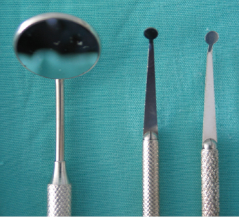
The modern approach to PRS is through magnification, illumination, and microsurgery. The literature is now reporting greatly improved prognosis when these techniques are employed. Surgical operating microscope (SOM) and endoscopy are the methods used today. Microsurgical instruments are used (Figs 12.1, 12.2 and 12.3). Far more detailed examination of the root apex is possible, allowing anatomic features such as isthmuses, accessory canals, fracture cracks, and crazing to be identified. Angled ultrasonic instruments can be used to prepare the retrograde cavity with the aid of micromirrors.
Stay updated, free dental videos. Join our Telegram channel
VIDEdental - Online dental courses


