Autotransplantation is a treatment alternative for replacing missing teeth, either those missing congenitally or those lost due to trauma or disease, by moving a tooth or a root to a more suitable position within the same individual.
Introduction
Teeth can be transplanted with good prognosis within the same individual. This is called autotransplantation and is defined as an organ or tissue moved from one location and placed in a different location within the same individual. Teeth can be transplanted from one place to another and a tooth or a root can also be transplanted to a more suitable position within the same socket.
Although replacement of lost or missing teeth in adults is nowadays more often carried out by implant treatment, there are a number of situations where autotransplantation seems to be a better alternative, especially in growing children and adolescents where implants should not be used due to the interference with growth of the alveolar process. A transplanted tooth with periodontal ligament (PDL) will follow and contribute to the development of the alveolar process and in many situations is therefore the first alternative for replacement in growing individuals. There are also some situations in adult patients where autotransplantation is a good alternative.
Donor Teeth
Transplants can be taken by using teeth in crowded regions or by strategic extraction when equalizing the number of teeth between quadrants. Moreover, many premolars are extracted as part of the orthodontic treatment and can be used as transplants. Third molars can also be used. The transplanted tooth must have a suitable root length and shape.
Teeth with roots under development are easier to extract and have a better prognosis. Developing teeth can revascularize while teeth with fully developed roots do not revascularize and have to be endodontically treated. It is also important to take into consideration the development and growth status of the individual and an interdisciplinary approach is important when planning.
Indications
Congenitally Missing Teeth
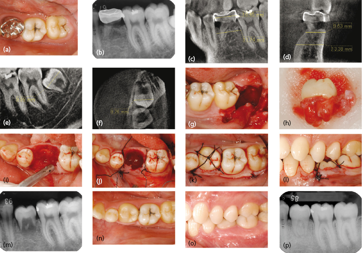
Tooth aplasia is a suitable indication for autotransplantation (Fig. 11.1). Donor teeth can be other erupted or impacted teeth or strategically extracted, e.g. taken from a crowded area or when a tooth does not have an antagonist. Another indication can also be to equalize teeth between quadrants. If the space is too small, preoperative orthodontic widening may be necessary. An advantage with this type of transplantation is that a natural biological condition for bone and soft tissue development of the alveolar process in the young growing patient is achieved and normal eruption is promoted.
Fig. 11.1 Transplantation of immature impacted third molar to the site of congenitally missing second premolar. (a) The patient is a 20-year-old male with a congenitally missing premolar 35 and a remaining deciduous molar 75. The root has been resorbed. (b) Radiograph showing resorption of 75. (c, d) Cone-beam CT images of the recipient site. The measurement of the recipient space has shown it is wide enough to accommodate the donor mesio-distally and bucco-lingually. (e, f) Cone-beam CT images of the donor tooth. The measurement has revealed the tooth is small enough to be placed into the recipient site. The lower left impacted third molar 38 is considered to be a good donor tooth for transplantation. (g, h) Extraction of the donor tooth 38. Note the Hertwig’s epithelial sheath is preserved at each apex. (i) Extraction of the tooth from the recipient site. (j) After careful try-in of the donor into the recipient site, suturing of the flap is performed without placing it in the socket. A knot is tied on the buccal aspect of the interproximal area and about 3 cm of one of the sutures is left. Another interrupted suture is tied across the vertical releasing incision on the mesial aspect. Again, 3 cm of one end is left. Next, a suture is placed through the interproximal area of the mesial aspect but in this procedure only one knot is tied loosely so that it allows space for the donor tooth to be placed into the socket. (k) Just after suture splinting. The loosely tied suture is tied tightly after placing the donor tooth. The tooth is tied into place by using a crisscross method. The remaining distal buccal end is tied with the mesio-lingual end and a knot is made on the center of the occlusal surface of the donor tooth. A horizontal mattress suture is tied on the disto-lingual aspect, and the remaining suture is tied to the remaining suture from the first crisscross knot. The remaining suture is then tied to the long remaining end of the suture from the vertical releasing portion. This allows the donor tooth to be seated in the correct position. Any excess suture is cut. (l) Occlusal adjustments are made on the antagonist so that articulating paper can freely slide through the contact. In this case, the opposing tooth was slightly extruded, so with the patient’s permission, some adjustments were made to the opposing tooth in contact. (m) Radiograph immediately after transplantation. Note the apices are wide open. (n, o, p) Follow-up after 1 year, 8 months. The transplanted tooth has erupted and is in contact with the antagonist. Pulp healing and root development are seen.
Unrestorable Teeth
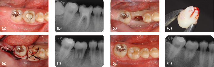
Autotransplantation may be indicated in situations with deep caries or where restoration or crown therapy is not feasible, or when endodontic treatment has failed (Fig. 11.2).
Fig. 11.2 Transplantation immature third molar 48 to replace an unrestorable first molar 46. (a) Occlusal view of the non-restorable 46. (b) Radiograph of 46. It was considered transplantation of an immature third molar would give a better prognosis than preserving 46 in this young patient. (c) 2 weeks after the extraction of 46. Transplantation was performed on the same day. (d) Extracted 48 as a donor. Note Hertwig’s epithelial sheaths are preserved apically. (e, f) Immediately after transplantation. (g, h) 4 years’ follow-up. The transplant shows a positive electric pulp test and normal healing is seen clinically a/>
Only gold members can continue reading. Log In or Register to continue