Principle
The fundamental principle of X-ray production is that X-rays are produced by the sudden deceleration or stoppage of rapidly moving stream of electrons at a positively charged metal target in a high vacuum tube.
Construction of x-ray tube
• The X-ray tube is like the heart of the X-ray generating system and is critical to the production of X-rays.
Leaded-Glass Housing
• The leaded-glass housing is a leaded-glass vacuum tube that prevents escape of X-rays in all directions.
• One central area of the leaded-glass tube has a “window” that permits the X-ray beam to exit the tube and directs it toward the aluminium discs, lead collimator and PID. It is also used for earthing.
Negatively Charged Cathode
• The cathode, or negative electrode, consists of two components:
a. Filament: Filament is a coiled wire made of tungsten, which produces electrons when heated.
b. Focusing cup: It is a cup-shaped holder made of molybdenum and houses the filament. It focuses the electrons in to a narrow beam and directs the beam across the tube toward the tungsten target of the anode.
• The purpose of the cathode is to supply the electrons necessary to generate X-rays. In the X-ray tube, the electrons produced in the negative cathode are accelerated toward the positive anode.
Positively Charged Anode
The rotating anode helps to dissipate heat and is mainly used in extraoral or cephalometric machines.
• The purpose of the anode is to convert electrons into X-ray photons and it consists of:
i. Is a wafer thin tungsten plate embedded in a copperstem. It serves as a focal spot and converts bombarding electrons into X-ray photons.
ii. The target is inclined at an angle of 20° to the central ray of electron to cause effective focal spot to be smaller in size (1 × 1 mm) in contrast to actual focal size (1 × 3 mm). This is known as “Line-Focus Principle”.
Sharpness of image increases by reducing the effective focal spot size. Copper stem: The copper stem dissipates the heat away from the tungsten target through conduction.
Circuits used in the production of x-rays
Transformers
Transformer is a device used to control voltage in the electrical circuit. Various transformers used in the production of X-rays are as follows:
• Step-down transformer: It has more coils in primary coil and reduces voltage from the incoming 110–220 line voltage to 3–4 V as required for filament circuit.
• Step-up transformer: It increases voltage from the incoming 110–220 line voltage to 65000–100000 V as required by a high-voltage circuit.
• Auto transformer: It serves as a voltage compensator that corrects the minor fluctuations in the current.
Timer
A timer completes the circuit with the high-voltage transformer and helps to control the time for which high voltage is applied to the tube.
Tube rating
The maximum safe intervals (seconds) the tube may be energized at a given range of voltage (kVp) and the tube current (mA) values is known as tube rating.
Duty cycle
Duty cycle related to the frequency with which successive exposures can be made.
Working of x-ray tube and production of x-rays
A series of steps involved in the production of X-rays are as follows:
• When the X-ray machine is turned on, the electric current enters the control panel from the wall outlet and travels to the tube head through the electrical wires in the extension arm.
• In the tube head, the current is directed to the filament circuit and the step-down transformer,which reduces the 110 or 220 entering-line voltage to 3 to 5 volts.
• The filament circuit uses the 3 to 5 volts to heat the tungsten filament in the cathode portion of the X-ray tube.
a. When the tungsten filament in the cathode is heated to incandescence or red hot, thermionic emission occurs.
b. Thermionic emission is defined as the release of electrons from the tungsten filament when the electrical current passes through it and heats the filament. The outer-shell electrons of the tungsten atom acquire enough energy to move away from the filament surface, and an electron cloud forms around the filament.
• The high-voltage circuit is activated when the exposure button is pushed. The electrons produced at the cathode are accelerated across the X-ray tube to the anode. The molybdenum cup in the cathode directs the electrons to the tungsten target in the anode.
• The electrons travel from the cathode towards the anode. When the electrons strike the tungsten target, their kinetic energy is converted to X-ray energy and heat.
a. Less than 1% of energy of electrons is converted to X-rays at anode and other 99% is lost as heat.
• The heat produced during the production of X-rays is carried away from the copper stem and absorbed by the insulating oil in the tube head.
• The X-rays produced are emitted from the target in all directions. The leaded-glass housing prevents the X-rays from escaping from the X-ray tube. A small number of X-rays are able to exit from the X-ray tube through the unleaded glass window portion of the tube.
• The X-rays travel through the unleaded glass window, the tubehead seal and the aluminium discs. The aluminium discs remove or filter the longer wavelength X-rays from the beam.
• The size of the X-ray beam is restricted by the lead collimator. The X-ray beam then travels down the lead-lined position indicating device (PID) and exits the tube head.
• The exposure time is the duration of time when X-rays are produced, it is about 0.8–0.9s.
• The X-ray tube does not emit a continuous stream of radiation, but a series of impulses of radiation. The number of impulses depends on the number of cycles per second in the electric current used. In a 60-s-cycle alternating current, there are 60 pulses of X-rays per second. Each impulse lasts only l/120 second as no X-rays are emitted in the negative half of the cycle when the polarity of the tube is reversed. A full-wave rectified X-ray machine produces 120 bursts of X-ray photons per second.
Production of X-rays is achieved by following two processes that are described as follows:
The Bremsstrahlung radiation accounts for most of the X-rays produced in dental machines,while characteristic radiation accounts for a very small part of X-rays produced.
Bremsstrahlung radiation
• Bremsstrahlung, is a German word for braking radiation. It is also called general radiation, white radiation or Brems radiation or breaking radiation.
• It is defined as X-ray radiation produced when high-speed electrons are suddenly stopped at the target. This process of rapidly decelerating the high-speed electron gives rise to Bremsstrahlung or braking radiation.
• Bremsstrahlung radiation is produced by either:
a. The electron directly hitting the nucleus of an atom of the target material or
b. Passage of the electron by the side or near the nucleus due to which the electron will be deflected or decelerated.
i. Electron directly hitting the nucleus. When the electron directly hits the nucleus of the tungsten atom in target material, the entire kinetic energy of it is transformed into a single X-ray photon.
ii. Numerically the energy of the resultant photon is equal to the energy of the electron, which is in turn equal to the kilo-voltage (kVp) applied across the X-ray tube.
a. When the electron comes closer to the nucleus
i. If the electron misses the hitting of nucleus and passes by the side of it, then the negatively charged high-speed electron is attracted towards the positively charged nucleus and decelerates thereby losing some kinetic energy, which is converted into X-ray photon.
ii. The electron that misses the nucleus continues to penetrate many such tungsten atoms before it imparts all its kinetic energy thus producing many low-energy X-ray photons. As a result Bremsstrahlung radiation consists of X-rays of many different energies and wavelengths and hence it is also called continuous spectrum.
Characteristic radiation
• When a high-speed electron dislodges the inner shell electron from the tungsten atom, it results in ionization of the atom. Once electron is dislodged, the remaining orbiting electrons rearrange to fill the vacancy; this produces a loss of energy that results in X-ray photon,with energy equal to the difference in the two orbital energy states. The X-ray thus produced is called characteristic radiation.
Q. 2. Define ideal radiograph and discuss the factors affecting the X-ray beam.
Or
Describe in detail the factors controlling x-ray beam.
Ans. According to HM Worth’s words, “An ideal radiograph is one that has desired density and overall blackness and which shows the part completely without distortion with maximum details and has the right amount of contrast to make the details fully apparent”.
Factors controlling the x-ray beam
The quality and quantity of the X-rays are controlled by various factors as described below:
Tube current
• The number of X-ray photons generated is determined by the tube current (mA).
• As the mA is increased, more number of electrons are generated at the cathode, which strike the target to produce more number of X-ray photons.
• The number of X-rays produced depends directly upon the number of electrons that strikes the target. The number of electrons is directly proportional to the tube current.
• Practically, the quantity of X-ray photons generated depends on both the mA and the duration of time the X-ray machine is operated.
Tube voltage
• Voltage is a measurement of force that refers to the potential difference between two electric charges. In simple terms, voltage is a measurement of electrical force that causes electrons to move from negative cathode to positive anode.
• Tube voltage controls the energy of electrons. As the kilovoltage peak (kVp) is increased, the energy of each electron striking the target increases resulting in increase in the number of X-ray photons generated.
• As kVp increases,there is an increase in
• As the kVp increases, the contrast of the resultant radiographic image decreases.
Exposure time
• Keeping mA and kVp constant,when the exposure time is doubled, the number of X-ray photons generated also doubles.
• The changes in the exposure time influences the quantity of X-rays produced.
• The effect of increasing or decreasing exposure time will control the quantity of X-ray photons.
• To compensate for the increased penetrating power of X-ray beam, when kVp is increased, an adjustment in exposure time is necessary.
Filtration
• An X-ray beam is composed of a spectrum of X-ray photons with different wavelengths and penetrating powers. Only those photons with sufficient energy and definite penetrating power contribute to image formation, whereas X-ray photons with less penetrating power will be absorbed by the soft tissues and cause unnecessary radiation exposure to the patient.
• Filtration is the process of removing X-ray photons of less penetrating power by placing a filter in the path of the primary beam, which allows only X-ray photons with sufficient energy to pass through.
• A filter is a device made up of an aluminium disc placed in the path of the primary X-ray beam to absorb X-ray photons of less penetrating power.
• Filtration is of three types:
a. Inherent filtration is produced by materials which the X-ray beam encounters as it leaves from the target. E.g.: The glass wall of the X-ray tube, insulating oil present around the tube, and the barrier material, which prevents the oil from leaking out. The inherent filtration usually provides 0.5 to 2.0 mm aluminium equivalent of filtration.
b. Added filtration: Added filtration refers to any additional aluminium disc placed in the path of the primary beam.
c. Total filtration: Total filtration means the sum of inherent and added filtration. The total filtration should be equivalent to 1.5 mm of aluminium upto 70 kVp and 2.5 mm of aluminium above 70 kVp.
• With the use of filters, the contrast and quality of film is increased, while the density is affected; therefore, when filtration is increased, a slight increase in exposure time is required.
Collimation
• Collimation is the process of restricting the size of the X-ray beam and thus the volume of the irradiated tissue of the patient from which the scattered photons originate.
• Collimator is a device that is used to shape or restrict the size of the X-ray beam striking the patient’s tissues.
• The collimator is made up of a material, which is capable of absorbing the radiation. For e.g., lead.
• Various collimators used in dental radiography are the diaphragm, tubular, and rectangular collimators.Among them, the rectangular collimators help in defining the X-ray beam to a size slightly larger than the size of the film.
Uses of collimation
Inverse square law
• Inverse square law states that the intensity of an X-ray beam at a given point is inversely proportional to the square of the distance from the source of radiation.
• The mathematical formula used to calculate inverse square law is given by
< ?xml:namespace prefix = "mml" />
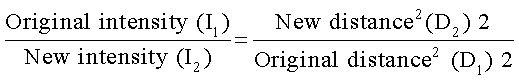
• The reason for this decrease in intensity of the X-ray beam is due to the divergent nature of the X-rays. If the distance from the source to the object is increased, the intensity of the X-ray beam decreases, thereby changing the image quality. E.g.: If the distance from the source to the film is doubled,say from 8 inches to 16 inches, it results in a beam that is one-fourth as intense.
Quality of the x-ray beam
• The quality of the X-ray beam refers to its mean energy or penetrating ability.
• X-rays with shorter wavelengths have more penetrating power, whereas those with longer wavelengths have less penetrating power and get absorbed by the patient’s soft tissues.
• The quality of an X-ray beam is governed by the kVp. When the kVp increases, it results in X-ray photons with high energy and better penetrating power.
Quantity of the x-ray beam
• Quantity of the X-ray beam refers to the number of X-ray photons produced.
• The amperage determines the electrons passing through the filament. When mA is increased, more number of electrons are released in the cathode and they strike the target to produce more number of X-ray photons.
• The quantity depends on the product of mA and exposure time in seconds (mAs).
Half-value layer
• Half-value layer (HVL) refers to the thickness of specified material such as aluminium required to reduce the intensity of an X-ray beam by one-half. Usually 2.0 mm filter is required in dentistry.
• Quality of X-ray beam can be determined by determining, its half-value layer (HVL). HVL is the useful way to designate the penetrating power of X-ray beam.
• HVL is the thickness of an absorber, usually aluminium, required to reduce the number of X-ray photons passing through it by one-half.
• Contrast and the quality of film is increased with the use of filters, while density is affected because increased filtration may result in absorption of some of the useful penetrating X-rays.
Stay updated, free dental videos. Join our Telegram channel
VIDEdental - Online dental courses


