Chapter 1
The oral cavity in health
INTRODUCTION
Before oral health educators (OHEs) can deliver dental health messages to patients, and confidently discuss oral care and disease with them, they will need a basic understanding of how the mouth develops in utero, the anatomy of the oral cavity (Figures 1.1, 1.2, 1.3 and 1.4) and how the following structures function within it:
Figure 1.1 Structure of the oral cavity (© Elsevier 2002. Reproduced with permission from Reference 1)
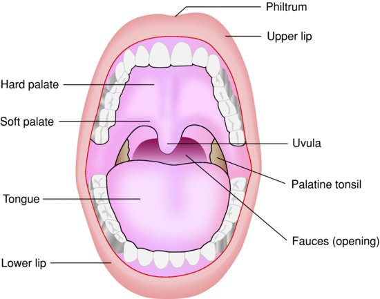
Figure 1.2 A healthy mouth, white person (© John Wiley & Sons, Ltd 2003. Reproduced with permission from Reference 2)
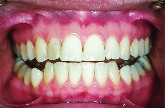
Figure 1.3 A healthy mouth, black person (source: Alison Chapman)
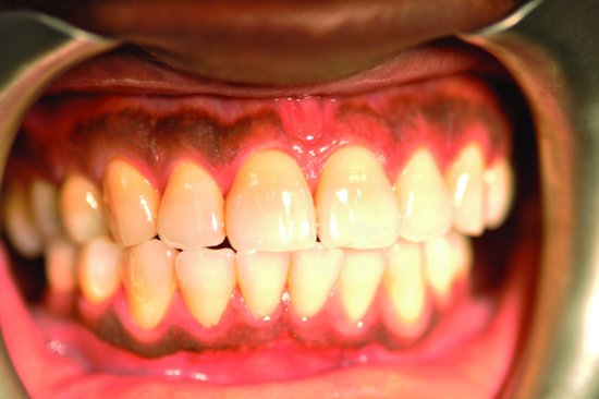
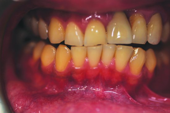
Figure 1.4 A healthy mouth, Asian person (source: Alison Chapman)
- Teeth (including dentition).
- Periodontium (the supporting structure of the tooth).
- Tongue.
- Salivary glands (and saliva).
ORAL EMBRYOLOGY
A basic understanding of the development of the face, oral cavity and jaws in the embryo and developing foetus will enable the OHE to discuss with patients certain oral manifestations of conditions that stem from in utero development (notably cleft lip and palate – Figure 1.5). (An embryo describes the growing organism up to 8 weeks in utero; a foetus describes the growing organism from 8 weeks in utero.)
Figure 1.5 Cleft lip (© iStockphoto.com/April Anderton)

Development of the face
At approximately week 4 in utero (Figure 1.6), the embryo begins to develop five facial processes (projections), which eventually form the face, oral cavity, palate and jaws by week 8 [3]:
Figure 1.6 Facial development at 4 weeks in utero (© John Wiley & Sons, Ltd. Reproduced with permission from Reference 3)
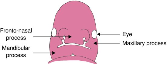
- Frontonasal process – forms the forehead, nose and philtrum (groove in upper lip).
- Maxillary process (two projections) – forms the middle face and upper lip.
- Mandibular process (two projections) – forms the mandible and lower lip.
Development of the palate and nasal cavities
Week 5
The frontonasal and maxillary processes begin to form the nose and maxilla. However, if the nasal and maxillary processes fail to fuse a cleft lip and palate will result [3]. A cleft lip can be anything from a small notch in the lip (incomplete cleft) to a wide gap that runs up to the nostril (complete cleft lip). One type of cleft palate (submucous) can be hidden.
There are two types of cleft lip:
- Unilateral – appears on one side of the lip at the philtrum.
- Bilateral – occurs on both sides of the lip, both sides of the philtrum.
Week 6
By week 6, the primary palate and nasal septum have developed. The septum divides the nasal cavity into two.
Week 8
By week 8, the palate is divided into oral and nasal cavities.
Development of the jaws (mandible and maxilla)
Week 6
By week 6, a band of dense fibrous tissue (Meckel’s cartilage) forms and provides the structure around which the mandible forms.
Week 7
By week 7, bone develops, outlining the body of the mandible, and as the bone grows backwards two secondary cartilages develop; these eventually become the condyle and coronoid processes. As the bone grows forward, the two sides are separated by a cartilage called the mandibular symphysis. The two sides will finally fuse into one bone approximately 2 years after birth. Upward growth of bone begins along the mandibular arch forming the alveolar process, which will go on to surround the developing tooth germs.
Week 8
By week 8, ossification (bone development) of the maxilla begins.
Tooth germ development in the foetus
Tooth germ (tissue mass) develops in three stages known as bud, cap and bell. The developing tooth germ can be affected by the mother’s health (see Chapter 20).
MAIN FUNCTIONS OF THE ORAL CAVITY
The oral cavity is uniquely designed to carry out two main functions:
TEETH
Different types of teeth are designed (shaped) to carry out different functions. For example, canines are sharp and pointed for gripping and tearing food, while molars have flatter surfaces for chewing. Tooth form in relation to function is known as morphology.
Dental nurses and health-care workers may remember from their elementary studies that there are two types of dentition (a term used to describe the type, number and arrangement of natural teeth):
Primary dentition
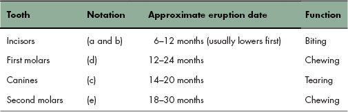
There are three types of deciduous teeth that make up the primary dentition (Figure 1.7): incisors, canines and molars (first and second). Table 1.1 details their notation (the code used by the dental profession to identify teeth), approximate eruption dates and functions.
Figure 1.7 Primary dentition (© Elsevier 2002. Reproduced with permission from Reference 1)
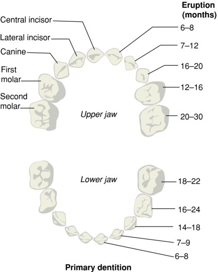
Table 1.1 Primary dentition (notation, approximate eruption dates and functions)
Secondary dentition
There are four types of permanent teeth that make up the secondary dentition (Figure 1.8): incisors, canines, premolars and molars. Table 1.2 details their notation, approximate exfoliation/eruption dates and functions.
Figure 1.8 Secondary dentition (© Elsevier 2002. Reproduced with permission from Reference 1)
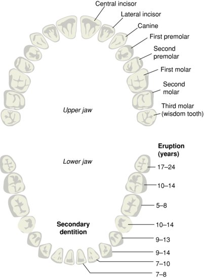
Table 1.2 Secondary dentition (notation, approximate exfoliation/eruption dates and functions)
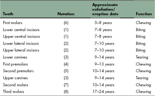
It is important to remember that these exfoliation/eruption dates are only approximate and vary considerably in children and adolescents. The educator should be prepared to answer questions from parents who are worried that their child’s teeth are not erupting at the same age as their friends’ teeth. Parents often do not realise, for example, that no teeth fall out to make room for the first permanent molars (sixes), which appear behind the deciduous molars.
Structure of the tooth
Tooth structure (Figure 1.9/>
Stay updated, free dental videos. Join our Telegram channel
VIDEdental - Online dental courses


