The oral cavity has the potential to be a major source of short-term and long-term complications from cancer therapy. Appropriate evaluation and elimination of potential sources of oral infection before cancer therapy is vital because oral bacteria are a known source of bacteremia and septicemia during cancer therapy. Cancer diagnosis with previous and planned treatment, past medical history, past dental history, current medications, drug allergies, social history, family history, laboratory values, extraoral findings, intraoral findings, and radiographic findings must all be evaluated in planning dental treatment for these complex cases.
The management of dental disease before and during cancer therapy poses many challenges to the dental practitioner. Cancer therapy has numerous potential short-term and long-term oral complications that may require modification of dental management strategy. Dental treatment may also require modification in situations where it must be delivered expeditiously and there is little time to institute an ideal treatment plan. For example, patients first diagnosed with acute leukemia begin induction chemotherapy within days of their diagnosis and will not have sufficient time for elective therapy and elimination of all sources of dental disease. During chemotherapy when white blood cell (WBC) counts are low, a dentist may elect to treat a dental infection with antibiotics rather than an extraction. In spite of this, pretherapy dental evaluation consisting of a good history, examination, and oral radiographs provide a baseline assessment that may be helpful if problems occur during cancer therapy.
Dental management of a patient in preparation for cancer therapy
The need for conducting a pretreatment oral evaluation depends on the cancer diagnosis and planned cancer therapy. Patients diagnosed with solid tumors (such as breast, prostate, lung, or colon cancers—the most common cancers in the United States) are not typically evaluated. Patients who are most frequently seen before chemotherapy or radiation are those who are at the highest risk for developing short-term and long-term complications as outlined above. These include the following:
- (1)
Patients undergoing chemotherapy followed by hematopoietic stem cell transplantation (HSCT) . Conditioning regimens for HSCT are generally myeloablative and place the patient at high risk for infectious complications during the period of pancytopenia. Autologous HSCT is used to treat lymphoma, multiple myeloma and some metastatic solid tumors to the marrow. In this procedure, the patient’s own stem cells are collected before conditioning, and then reinfused to reconstitute the marrow that has been ablated by chemotherapy. Allogeneic HSCT is generally performed in patients with leukemia or bone marrow failure syndromes such as aplastic anemia where the diseased marrow is replaced by healthy marrow from a donor. Following allogeneic HSCT, patients may be immunosuppressed in the long term because of prophylaxis for chronic graft-versus-host disease (GVHD), slow immune reconstitution, and treatment for chronic GVHD.
- (2)
Patients who require head and neck radiation therapy. Head and neck cancers that are typically treated with head and neck radiation therapy include squamous cell carcinoma, salivary gland malignancies, and lymphoma. Radiation at therapeutic doses induces long-term irreversible damage to the salivary glands, connective tissues, vasculature, and healing potential of the jawbones, and in particular the mandible.
The following are the goals of dental management before the start of cancer therapy.
Eliminate or stabilize oral disease to minimize local and systemic infection during and after cancer therapy
Short term
Cytoreductive therapy generally leads to low WBC counts and in particular low neutrophil counts (neutropenia), which increases susceptibility to infection, in particular bacterial infections. Since most odontogenic infections such as caries, periodontal infection and third molar infections are usually of bacterial origin, elimination of potential sources of infection from the oral cavity is a key strategy to prevent new infections or exacerbation of existing chronic infections . Ideally, all patients should be returned to a stable, if not perfect state of dental health before cytoreductive therapy.
Mucosal injury or mucositis is a common side effect of cancer therapy and removal of sharp edges of teeth or restorations may help to reduce trauma to the mucosa and reduce the severity of mucositis and attendant pain and discomfort. Furthermore, ulcers of mucositis may act as a gateway for ingress of oral bacteria in profoundly myelosuppressed individuals with the potential for bacteremia and septicemia . The frequency of viridans streptococci in neutropenic patients has become more common, with Streptococcus mitis being the most common species identified . Up to one third of viridans streptococci–infected patients can develop shock syndrome .
Long term
Radiation therapy to the head and neck has the potential for increased caries risk and reduced healing capacity (especially of the bone) in the long term . Elimination of dental disease by judicious restorative dentistry and periodontal treatments, and extraction of teeth with questionable prognosis are important preventive strategies to avoid future dental extractions, an important risk factor for postradiation osteonecrosis. Custom trays for prescription fluoride applications are often fabricated for patients since lifelong fluoride therapy helps to minimize radiation-induced dental caries. Patients treated with intravenous (IV) bisphosphonates are at risk for developing bisphosphonate-related osteonecrosis of the jaws . Because invasive dental procedures such as extractions are a risk factor for this condition, elimination of dental disease to reduce the necessity for such dental procedures in the long term is an important aspect of patient management.
Patients who undergo allogeneic hematopoietic stem-cell transplantation may develop chronic GVHD (see the article by Schubert and Correa elsewhere in this issue). This disease may result in painful mucosal ulcerations, increased caries rates, hyposalivation, and sometimes fibrosis, limiting mouth opening. Similar to patients with radiation-induced salivary gland hypofunction, caries even when incipient must be treated and dental treatment should not be deferred.
Identify issues specific to the cancer diagnosis
A thorough examination of the mouth for oral involvement of the primary tumor such as leukemic infiltrates (especially in the gingiva) and jaw involvement from multiple myeloma should be performed. Such oral involvement that may not have been noted previously may change the stage of the patient’s disease and impact future cancer therapy.
Educate the patient regarding short-term and long-term oral complications from cancer therapy
This provides an opportunity to educate the patient about the role of dental health in systemic disease. Patients should be informed about why a dental evaluation is important before cancer therapy, what to expect during cancer therapy (such as mucositis and xerostomia), and measures that can be taken to minimize side effects of therapy. The importance of long-term follow-up should also be stressed, especially with respect to postradiation caries and osteonecrosis, bisphosphonate-related osteonecrosis of the jaws, and chronic GVHD in the appropriate patient population. Written information is usually helpful for patients and many cancer centers provide such information for patient education. The National Institutes of Health provides information and pamphlets free of charge at the following Web site: https://cissecure.nci.nih.gov/ncipubs/searchResults.asp?subject2=Coping+with+Cancer .
Pretreatment evaluation and treatment planning
The pretreatment evaluation for patients with cancer requires a thorough understanding of the cancer diagnosis and stage, and should include a review of systems, current medications, drug allergies, and social and family history, in addition to a comprehensive oral examination ( Box 1 ). Communication with the patient’s oncologist with regard to previous and planned cancer therapy helps the dentist and dental specialist to plan and schedule dental treatment appropriately.
-
Review recent medical history
-
Past medical history
-
Medications
-
Drug allergies
-
-
Social history
-
Family history
-
Review current cancer diagnosis
-
Previous cancer treatment
-
Planned cancer treatment
-
-
Past dental history
-
Frequency of past visits, including hygiene
-
History of extractions, periodontal surgery, endodontics, pain, swelling, bleeding, ulcers
-
-
Present oral complaint
-
Character, location, onset, radiation, intensity, duration, and exacerbating factors of the complaint
-
Management of current oral complaints
-
-
Oral examination
-
Extraoral
-
Observe for asymmetries, swelling, or skin lesions
-
Palpate for nodes, salivary glands, temporomandibular joint, muscles of mastication, and neck muscles
-
-
Intraoral
-
Remove appliances: examine for fit, function, and aesthetics
-
Evaluate for soft tissue abnormalities
-
Examine teeth for plaque, calculus, caries, mobility, faulty restorations, fractures
-
-
-
Radiographs
-
Panoramic radiograph and full-mouth radiographic series
-
Examine for bone loss, caries, root tips, impactions, calculus, periapical/furcation radiolucencies, fractures
-
-
Laboratory values
-
Complete blood count + WBC differential
-
prothrombin time/international normalized ratio/partial thromboplastin time
-
The logistics of a pretreatment evaluation can be complicated by many factors. For patients who prefer to be treated by their own dentist or who may not be able to travel to the cancer center for dental treatment, an off-site dental evaluation program may be used . In this type of program, the dentist performs the entire evaluation and treatment planning guided by written instructions from the dental oncology service. All treatment is provided by the dentist or dental specialist in the community. Although this has been used primarily for patients scheduled for HSCT, the same program may be used for any of the other patient populations discussed above. Off-site evaluations save time, are preferred by many patients, save hospital resources, and are well-accepted by dentists in the community who welcome the opportunity to play an important role in their patients’ cancer care.
Presenting symptoms
Cancer patients can present with a wide range of oral symptoms and a thorough review of the CHaracter, Location, Onset, Radiation, Intensity, Duration, and Exacerbating factors (CHLORIDE is a useful mnemonic) of these symptoms helps the dentist arrive at a diagnosis. Distinguishing between acute infections of pulpal versus periodontal origin, or temporomandibular joint (TMJ) versus odontogenic origin is vital in arriving at an appropriate treatment plan. Importantly, patients may experience pain directly related to their cancer. Patients with squamous cell carcinoma in the oral cavity often experience pain from the tumor and this must be differentiated from pain of odontogenic origin .
A history of extractions, periodontal surgery, and endodontic therapy together with a history of previous pain, swelling, or bleeding should be obtained in case these areas become symptomatic during cancer therapy.
Laboratory values
A recent complete blood count (CBC) must be obtained on patients with leukemia, myeloma, and marrow failure syndromes such as aplastic anemia before examination of the patient. The CBC should be evaluated in the context of the patient’s disease. A high neutrophil count in a patient with acute myeloid leukemia does not necessarily indicate an infection, but is typical of the disease. Since many of these neutrophils are not functional, the patients will still be highly susceptible to infection. Marrow involvement by primary malignancy such as leukemia or metastatic disease reduces the volume of “normal” hematopoietic elements resulting in neutropenia, thrombocytopenia, and anemia .
Necessity for antibiotic prophylaxis
Another consideration for cancer patients is the concern regarding infections with indwelling catheters such as Hickman lines. The rate of bloodstream infection with Hickman catheters has been reported as 4.7 episodes per 1000 catheter-days , with neutropenia an important risk factor for catheter-related infections . A Cochrane review of antimicrobial prophylactic antibiotics before insertion of long-term tunneled central venous catheters in oncology patients found administration of an antibiotic before catheter insertion did not significantly decrease catheter infection, but that flushing the catheter with vancomycin and heparin did have a positive overall effect . There is no guideline regarding the necessity for antibiotic prophylaxis before invasive oral procedures as there is no clear link between dental procedure–related bacteremia and catheter infection. However, antibiotic prophylaxis before invasive oral procedures may be recommended for patients with central venous catheters; the current American Heart Association protocol for prevention of infective endocarditis following oral procedures is frequently used for these patients.
Clinical examination
Patients with squamous cell carcinoma presenting for preradiation therapy evaluation may have healing surgical sites both extra- and intraorally. The clinician must be empathetic in the presence of oral discomfort and take care to minimize trauma to the soft tissues during the examination.
Extraoral examination
The head and neck examination should consist of an inspection for asymmetries, swellings, or skin lesions, followed by palpation of the lymph nodes of the submental, submandibular, and cervical chains. Lymph nodes should be evaluated for the presence of pain to palpation, mobility, or fixation. A lymph node that is fixed and nonpainful is a common finding in metastatic tumors to the node while a freely movable and tender node is more likely to represent an infectious or inflammatory process. Salivary glands, the temporomandibular joint (TMJ) area, muscles of mastication, and neck muscles should be palpated to provide baseline information.
Intraoral examination
A systematic approach to the oral cavity is important to avoid missing more subtle pathologic processes. The soft tissues of the buccal mucosa, floor of mouth, tongue, palate, and oropharynx should be examined for erythema, ulceration, erosions, mucosal hemorrhages, swellings, or other lesions. The saliva expressed from the major salivary glands should be examined for presence, color, and consistency. The presence of cloudy and thick saliva may represent a chronic bacterial infection of the salivary gland(s).
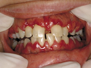
Patients with leukemia, especially acute monocytic or myelomonocytic leukemia may present with leukemic infiltration of the oral cavity . These patients may have boggy, swollen gingiva that bleed when brushing or upon palpation during the clinical examination ( Fig. 1 ). Petechiae and ecchymoses may develop with decreased platelet counts (thrombocytopenia) .