Figure 2.7 (a–d) Jaw deviates to the left at 23 mm opening and then centers again on maximum mouth opening (45 mm).
G. Intraoral Examination
- Marginal gingivitis and presence of plaque.
- Class I molar and canine relationship on the right side and class II on the left side, with asymmetrical incisor midline.
H. Additional Examinations and Findings
- Panoramic radiograph: normal findings (Figure 2.8). MRI confirmed the clinical diagnosis of left TMJ disc displacement with reduction, with the disc located ahead of the condyle in the closed mouth position (Figure 2.9a) and physiologically positioned over the condyle, showing a “butterfly” form, in the opened mouth position (Figure 2.9b). No effusion could be detected (Figure 2.9c). The right side showed a disc in normal position in both the closed (Figure 2.9d) and opened mouth position (Figure 2.9e).
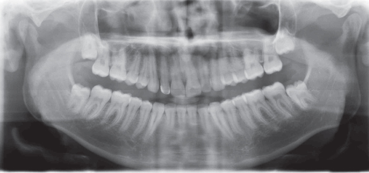
Figure 2.8 Panoramic radiograph showing an absence of gross morphological changes of the TMJ condyles.
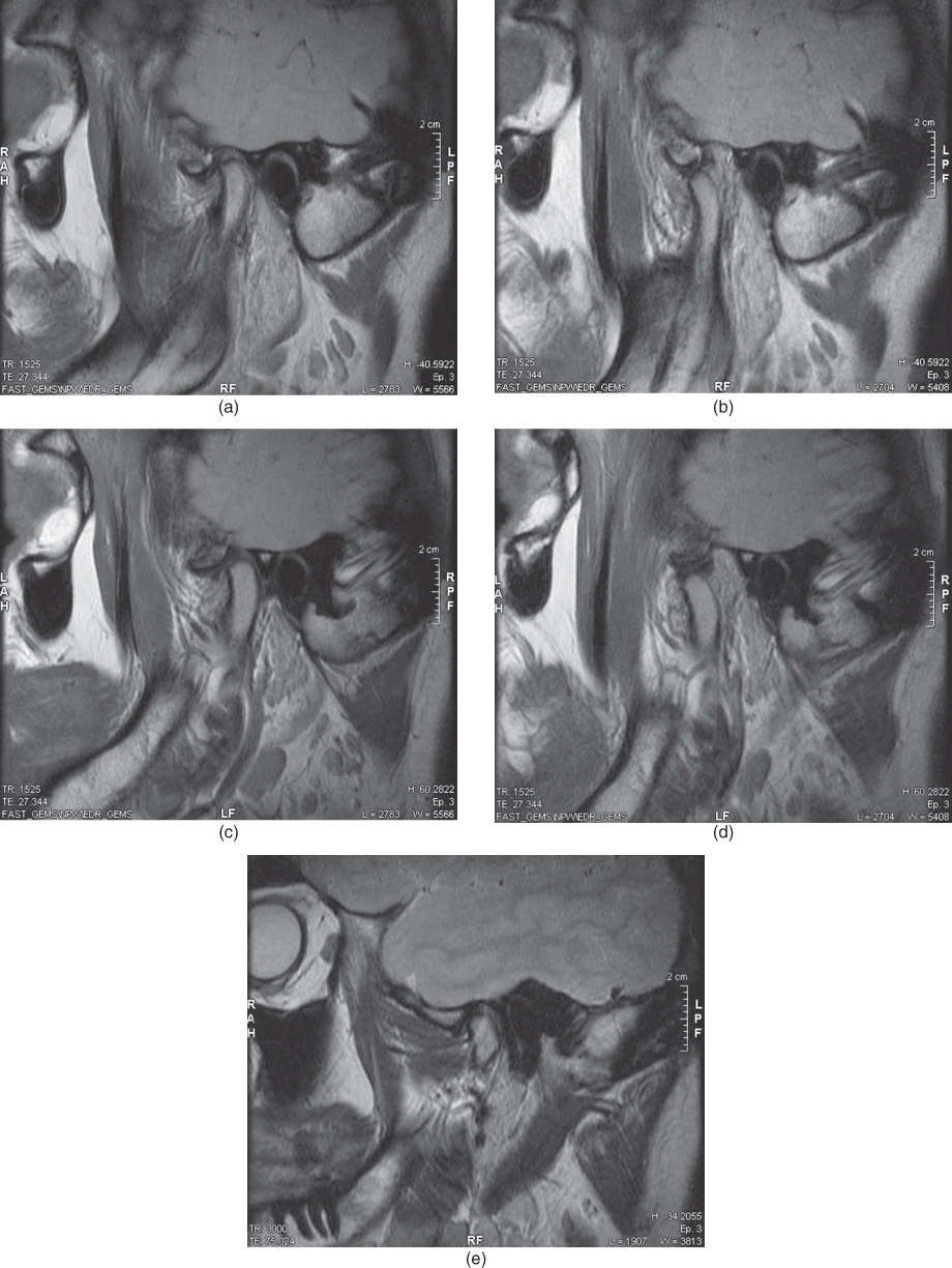
Figure 2.9 MRI showing disc displacement with reduction in the left TMJ (a, b) with absence of TMJ effusion on T2-weighted image (c) and normal disc positions in the right TMJ (d, e).
I. Diagnosis/Diagnoses
DC/TMD
- Disc displacement with reduction (left side).
J. Case Assessment
- Patient with a nonpainful disc displacement in the left TMJ. No functional interference. In the case presented here there was no need to get deeper into the psychosocial assessment for prognostic purposes owing to the absence of pain and, consequently, pain-related disability.
K. Evidence-based Treatment Plan including Aims
- Counselling and reassurance. However, the patient may be worried by the click sounds or misinformed about their relevance. That is why it is fundamental to spend enough time to reassure patients during the first appointment to increase the patient’s knowledge, correct the expectations and to lower any anxiety. This includes showing a drawing of the TMJ and the jaw muscles, explaining how the TMJ works and what a disc displacement is. This should also include information that this is a benign condition that may very well fluctuate over time.
L. Prognosis and Discussion
- Absence of pain and pain-related disability (GCPS grade 0) strongly indicate an observational approach.
- The prognosis regarding escalating symptoms is good. There is no increased risk of developing a disc displacement without reduction in this case, compared with a normal joint case.
Background Information
- Disc displacement with reduction is an intracapsular biomechanical disorder involving the condyle–disc complex. In the closed mouth position the disc is in an anterior position relative to the condylar head and the disc reduces upon opening of the mouth. Displacements can be anterior, medial, lateral, and posterior, with the antero-medial one being the most common direction. Clicking, popping, or snapping noises may occur with disc reduction.
- In the closed mouth position, the TMJ disc is in an anterior position relative to the condyle within the glenoid fossa. During jaw opening the disc is “recaptured” by the condyle, and in the maximum opening position it is located with its intermediate band positioned between the condylar apex and the articular tubercle. The recapture of the disc during mouth opening produces a click sound, which is often audible also during jaw closing movement (i.e., reciprocal click). The click sound during jaw closing is due to the disc losing its correct relationship with the condyle, and is usually heard at a lower interincisal opening distance. A differential diagnosis should be considered with bone changes (e.g., osteophytes; deviations in form) producing a sound at the same interincisal distance every time, due to the fixed obstacle.
(De Leeuw et al., 2013; Schiffman et al., 2014)
Stay updated, free dental videos. Join our Telegram channel
VIDEdental - Online dental courses


