Introduction
Our objective was to compare vertical alveolar growth in areas adjacent to infraoccluded deciduous molars with growth in areas of deciduous molars and normal occlusion for a period of at least 1 year by using digital subtraction radiography.
Methods
This case-control study included 40 pairs of panoramic radiographs of growing patients with infraoccluded deciduous molars and 40 pairs of radiographs of patients without infraoccluded deciduous molars. One radiograph at baseline was obtained at diagnosis, and the other at least 1 year later. The subjects and the controls were matched according to chronologic age and time interval between the 2 radiographs. The 2 groups were compared with regard to vertical alveolar growth and vertical tooth movement. Measurements were assessed by using nonparametric tests (Mann-Whitney and Friedman) and a multiple comparison test. Significance was set at 5%.
Results
A statistically significant difference was observed between the groups with regard to vertical alveolar growth measured on the bone crest between the first permanent molars and second premolars.
Conclusions
Vertical alveolar growth between the first permanent molar and the second premolar adjacent to the infraoccluded teeth was smaller than in areas adjacent to teeth with normal occlusion.
Dental ankylosis, also called teeth in infraocclusion, is commonly found in deciduous and transitional dentitions, with a prevalence ranging from 8% to 14% in patients aged 6 to 11 years; severity is usually mild (61.3%) or moderate (30.4%). Deciduous teeth are more commonly affected than permanent teeth, especially the first and second deciduous molars.
When there is infraocclusion, growth and development of the alveolar bone are affected, with a consequent reduction in bone height, thus precluding eruption of the affected tooth that remains in infraocclusion. Early diagnosis of the infraocclusion is essential for the establishment of effective preventive measures or treatment planning, including invasive procedures, always associated with adequate follow-up.
Radiographic investigation is often recognized as an important diagnostic method in the follow-up of patients with infraoccluded teeth. It allows detection of an ankylosed area based on the absence of continuity of the periodontal ligament in the region where the cementum fuses with the alveolar bone. Moreover, digital subtraction of panoramic radiographs has been successfully used to evaluate the behavior of implants, periapical lesions, and condylar alterations. This method allows evaluation of small changes by the superimposition of 2 radiographs, as shown in previous studies.
No scientific evidence or research data are available confirming the relationship between infraocclusion of deciduous molars and vertical alveolar growth abnormalities in areas adjacent to the affected teeth. Therefore, the objective of this study was to compare vertical alveolar growth in areas adjacent to infraoccluded deciduous molars with areas of deciduous molars with normal occlusion for at least 1 year, based on measurements obtained with digital subtraction radiography.
Material and methods
The study protocol was approved by the Research Ethics Committee at Universidade Federal do Rio Grande do Sul in Brazil (protocol number 05/08). Pairs of panoramic radiographs of 80 patients in the transitional dentition, aged 6 to 9 years, and with the first permanent molars having at least one third of root formation, were selected from the files in the Department of Orthodontics and Pediatric Dentistry of the School of Dentistry. Half of the radiographic image pairs (n = 40) were obtained from patients who had infraoccluded deciduous molars (group 1, subjects) and the other half (n = 40) from age-matched patients without infraocclusion (group 2, controls).
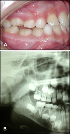
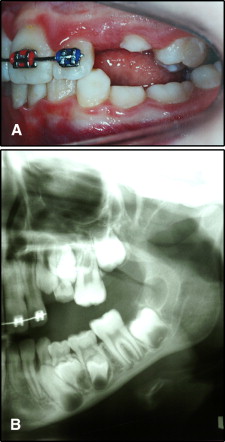
Inclusion criteria for group 1 were infraocclusion affecting at least 1 mandibular deciduous molar and the availability of 2 panoramic radiographs obtained within a time interval of at least 12 months, one at diagnosis (baseline, T1) and the other at least 1 year later (T2). After initial clinical examinations of the patients by 2 experienced examiners (a pedodontist [C.D.] and an orthodontist [L.C.]) for submergence of the deciduous molars, intraoral photographs and study models were evaluated to diagnose infraoccluded second deciduous molars. Infraocclusion was considered to be mild when the occlusal surface of the tooth was approximately 1 mm below the occlusal plane (line drawn between the first molar and the canine) and moderate when the occlusal surface of the tooth and both marginal crests were at the same level or slightly below the contact point with the adjacent teeth ( Figs 1 and 2 ). Measurements were recorded to the nearest tenth of a millimeter with a digital caliper (Mitutoyo Digimatic, Mitutoyo, Hampshire, United Kingdom). Two independent investigators (C.D. and L.C.) made space measurements, and the average of the 2 measurements was used in the analysis. The sample included 18 teeth with moderate infraocclusion and 22 with mild infraocclusion.
Patients undergoing treatment with systemic medications or with a history of growth disorders, such as gigantism, nanism (abnormally small size or stature), cleidocranial dysostosis, and ectodermal dysplasia, among others, were excluded from the sample. Patients needed to have their mandibular first permanent molars fully erupted and normally shaped. Deciduous molars had to have at least a third of their root length with no mobility. Subjects with dental anomalies of number and form in the deciduous dentition were also excluded from the sample.
The control patients were matched to each subject based on chronologic age and time interval between the T1 and T2 radiographs.
The mean ages were 8.0 ± 1.2 years in group 1 and 8.1 ± 1.2 years in group 2. The mean time intervals between the 2 radiographs comprising the pairs were 27.3 ± 17.9 months in group 1 and 25.8 ± 13.4 months in group 2.
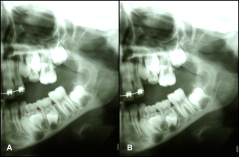
The radiographs were digitized with a resolution of 150 dpi at 8 bits and saved as jpg files, divided into left and right sides. Measurement points were marked on the digital copies of the T1 and T2 images by a previously trained examiner (C.D.). For the assessment of vertical tooth movement, the cusp tips of the first permanent molar, second premolar, first premolar, and permanent canine were marked with points ( Fig 3 , A ). For the assessment of vertical alveolar growth, lines were marked along the crest of the ridge from the permanent first molar up to the permanent canine of each hemi-arch in each image ( Fig 3 , B ).
Each radiographic image pair was assessed by the same examiner (C.D.), previously calibrated in a pilot study. Briefly, 10 images were selected, superimposed, and measured 3 times by the same examiner. The intraclass correlation coefficient showed excellent agreement among the 3 measurements (r i = 0.947; P <0.001).
With the digital subtraction radiography method and Photoshop (version 7.0; Adobe, San Jose, Calif), the 2 images obtained from each patient at T1 and T2 were superimposed ( Fig 4 ). Vertical and horizontal adjustments (sliding and rotating 1 image on another) were made based on the cortical border of the mandible and the mesiodistal diameter of the first permanent molar.
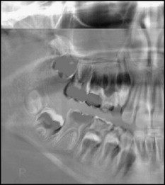
Vertical tooth movement and vertical alveolar growth were measured in millimeters, to an accuracy of 0.1 mm, by using the digital ruler tool in Adobe Photoshop, as the distance between the points previously marked on the cusp tips and on the bone crest of each image, respectively. Growth and movement were expressed as the difference in the distance between the reference points marked on the T1 and T2 images. Measurements made after superimposition of the 2 images aimed to prevent possible errors in the measurements of the 2 images of the same patient.
Statistical analysis
Measurements obtained in the 2 groups were assessed and compared by using nonparametric tests (Mann-Whitney and Friedman), as well as a multiple comparison test. Nonparametric tests are used for small samples and are not based on normal distribution. The Mann-Whitney test, a nonparametric counterpart to the independent samples t test, was used when at least 1 ordinal dependent variable was not assumed to be normally distributed. The Friedman test was used for independent variables with 2 or more levels and when at least 1 dependent variable was not assumed to be normally distributed. Significance was set at 5%.
Results
The Table shows the results from groups 1 and 2. There were no statistically significant differences between the groups with regard to vertical tooth movement. However, in the assessment of vertical alveolar growth, a statistically significant difference was observed in the area of the first permanent molar and second premolar.
| Measurement and site | Group 1 (subjects) | Group 2 (controls) | P | ||||||
|---|---|---|---|---|---|---|---|---|---|
| Median | 25th percentile | 75th percentile | Mean rank | Median | 25th percentile | 75th percentile | Mean rank | ||
| Vertical tooth movement | |||||||||
| First molar | 2.4 | 1.5 | 3.6 | 1.22 c | 3.0 | 1.6 | 4.2 | 1.22 d | 0.184 |
| Second premolar | 6.7 | 4.5 | 9.3 | 2.28 b | 6.6 | 4.2 | 9.4 | 2.19 c | 0.903 |
| First premolar | 9.3 | 5.3 | 12.3 | 2.74 b | 10.3 | 5.6 | 16.4 | 2.88 b | 0.167 |
| Canine | 14.4 | 10.3 | 21.8 | 3.76 a | 13.9 | 8.1 | 23.3 | 3.72 a | 0.652 |
| Vertical alveolar growth | |||||||||
| Distal molar | 2.4 | 1.9 | 2.9 | 2.51 a | 2.6 | 1.6 | 4.7 | 2.71 a | 0.111 |
| Between first molar and second premolar | 2.0 | 1.2 | 2.9 | 2.28 a | 2.4 | 1.9 | 4.0 | 2.55 a | 0.009 ∗ |
| Between second premolar and first premolar | 2.1 | 1.5 | 3.4 | 2.43 a | 2.4 | 1.3 | 4.0 | 2.34 a | 0.404 |
| Mesial first premolar | 2.7 | 1.6 | 3.8 | 2.79 a | 2.4 | 1.5 | 3.9 | 2.40 a | 0.733 |
Stay updated, free dental videos. Join our Telegram channel
VIDEdental - Online dental courses


