Abstract
The aim of this study was to evaluate the safety, utility and morbidity associated with the treatment of mandibular subcondylar fractures using the retromandibular transparotid approach and to evaluate the stability of a single 2 mm miniplate fixation system for such fractures. Forty-two cases with 48 mandibular subcondylar fractures were analysed prospectively for 12 months and evaluated for functional results, scar, postoperative complications and stability of fixation. There were three cases of suboptimal occlusal status, two cases of haematoma that were drained and resolved, eight patients with facial nerve weakness which resolved in a few weeks, and three cases of salivary fistulae that resolved after treatment. All cases showed stable osteosyntheses. Maximal postoperative interincisal distance was 32–61 mm (mean 44 mm). Four patients had deflection on opening, while clicking on opening or chewing was observed in five patients. The postoperative scars were well accepted by all patients. The results of this study suggest that a retromandibular approach will facilitate accurate reduction and fixation of subcondylar fragments with a good cosmetic result and minimal complications. A single 2 mm miniplate fixation provides stable results.
Fractures of the mandibular condyle are common and account for 25–35% of all mandibular fractures reported in the literature . Their management remains a controversial issue. Traditionally, they have been treated with intermaxillary fixation followed by physiotherapy, but there is evidence of functional disharmony and compromised results in a significant percentage of adult patients treated with closed treatment . A clinical investigation of 52 patients concluded that low subcondylar fractures in adults, treated with closed methods, resulted in complications . These included malocclusion, mandibular asymmetry, restricted masticatory function, malunion or nonunion of fragments, disc displacement, ankylosis and pain on the affected side. In the same study, a group of patients treated with open reduction had minimal complications. This was attributed to precise re-alignment of the fractured segments, which would otherwise be difficult or impossible via closed treatment techniques. The function of the lateral pterygoid muscle tends to displace the condylar segment in an anterior-medial direction, and there is no other structure to counteract the force and the direction of this vector. Simple manipulation of the distal fragment, including distraction, does not aid in the proper reduction of the proximal segment. The condylar head frequently remains displaced and further functional disharmonies are to be anticipated. Rehabilitation is quicker with surgical treatment, enabling the temporomandibular joint and muscles of mastication to function normally faster . Even with a consensus developing on the preference for open reduction and internal fixation of these fractures , the clinician is still faced with the dilemma about an optimal approach to the ramus-condylar unit. The various approaches that have been published stand testimony to the shortcomings of most techniques . Limited access and injury to the facial nerve are the most common problems. With the development of improved surgical techniques and the acceptance of rigid fixation, functional reduction can be achieved readily.
Pre- and post-auricular approaches are suitable for intracapsular and high condylar neck fractures where open reduction with internal fixation (ORIF) is indicated . Submandibular, retromandibular and the rhytidectomy modification approaches are preferred for low condylar neck fractures . The intraoral approach and its endoscopically assisted modifications may offer better cosmetic results . A coronal approach can provide access if there are other indications to use this incision. The authors’ preference for low condylar neck fractures, when ORIF is indicated, is miniplate and screw fixation via the retromandibular approach. This technique was first described in 1967 in relation to vertical subcondylar osteotomies and was later popularized for the management of condylar fractures .
This prospective clinical study was carried out on 42 patients to assess the duration, efficacy, stability and safety of surgical treatment of extracapsular displaced/dislocated condylar fractures with the retromandibular approach using a single 2 mm miniplate system and to describe the complications encountered.
Patients and methods
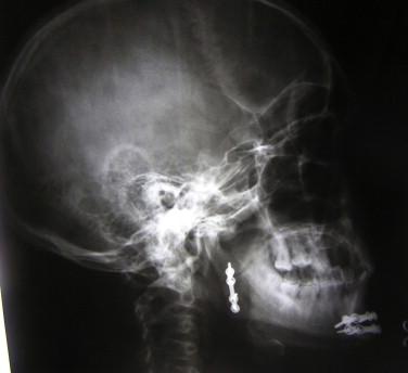
From June 2006 until June 2010, 42 patients with 48 extracapsular displaced/dislocated condylar fractures were treated with open reduction and internal fixation using a 2 mm miniplate and screws. All patients gave written informed consent and an institutional ethical committee approved the study. After taking the patient’s history and physical examination, radiological imaging of the mandibular ramus and condyle was performed using any combination of orthopantomogram ( Fig. 1 ), Water’s view, lateral cephalogram, posteroanterior/anteroposterior view of mandible or computed tomography. In cases where patients had additional radiographs at the time of presentation, they were used for diagnosis. Using any combination of these imaging methods, complete information about the level and type of fracture, as well as the degree and direction of displacement in all three dimensions was obtained.

All patients were intubated via the nasotracheal route and underwent maxillomandibular fixation with Erich arch bar and elastics placed at the beginning of the procedure. An incision 3–5 cm in length, parallel and posterior to the posterior border of the mandible was made starting 0.5 cm below the ear lobe ( Fig. 2 ) . The parotid capsule was identified after dissection through skin, subcutaneous fat and platysma. The parotid capsule was incised and blunt dissection performed with a curved haemostat to expose the masseter muscle. Facial nerve branches, if encountered, were carefully dissected out for a short distance with the help of a nerve stimulator and retracted either superiorly or inferiorly. The pterygomassetric sling at the posterior border of the mandible was incised with a 6–8 cm incision and further dissection was carried out subperiosteally after incising the periosteum at the posterior border of the mandible to the sigmoid notch exposing the fracture site. The condylar fracture was subsequently reduced and fixed using one 2 mm titanium miniplate (four holes with bridge) and four screws (2 mm × 8 mm) ( Fig. 3 ). The pterygomassetric sling and the parotid capsule were closed in two layers using 4-0 polyglactic acid sutures (Vicryl, Johnson and Johnson, India Ltd.). Skin was closed using 5-0 nylon sutures (Ethilon, Johnson and Johnson, India Ltd.). Maxillomandibular fixation was released at the end of the procedure. A note was made of the time taken to perform the procedure.


Postoperatively, patients were recommended to have a soft diet for 4 weeks. They were immediately encouraged to practice mouth opening and closing. Radiological imaging was performed, using the same views as preoperatively ( Fig. 4 ). Patients were usually discharged 3–5 days postoperatively. Sutures were removed 7 days postoperatively. A note was made of all intraoperative and early or late complications following the procedure, such as facial nerve palsy, salivary fistula, auricular anaesthesia, wound infection, haematoma formation, scar and miniplate fracture. Subsequently, patients had regular follow-up checks at 1 week, and 1, 3, 6 and 12 months postoperatively. Clinical examination was carried out by an independent examiner who was a physician regularly working with trauma patients and included factors such as pain and fatigue during mandibular mobility, audible clicking or crepitus in the affected joint, maximal interincisal opening, lateral deflection, occlusal status compared with pretraumatic occlusion, joint tenderness (at rest, on opening, loading or palpation), facial nerve function, ear sensitivity and scar. Postoperative facial nerve function was assessed using the House–Brackman Grading Scale ( Table 1 ) . Scar assessment was made using the Patient and Observer Assessment Scale ( Table 2 ) . Patients were asked to state their overall satisfaction regarding the course and outcome of treatment. The relationship between variables within treatment groups was assessed using Spearman’s rank correlation coefficients. Statistical significance was defined at P < 0.05.
| Grade | Description | Characteristics |
|---|---|---|
| I | Normal | Normal facial function in all nerve branches. |
| II | Slight | • Gross: Slight weakness on close inspection, slight synkinesis. |
| • At Rest: Normal tone and symmetry. | ||
| • Motion: Forehead: Good to moderate movement. Eye: Complete closure with minimum effort. Mouth: Slight asymmetry. | ||
| III | Moderate | • Gross: Obvious but not disfiguring facial asymmetry. Synkinesis is noticeable but not severe. May have hemifacial spasm or contracture. |
| • At Rest: Normal tone and symmetry. | ||
| • Motion: Forehead: Slight to moderate movement. Eye: Complete closure with effort. Mouth: Slight weakness with maximum effort. | ||
| IV | Moderately Severe | • Gross: Asymmetry is disfiguring and/or obvious facial weakness. |
| • At Rest: Normal tone and symmetry. | ||
| • Motion: Forehead: No movement. Eye: Incomplete eye closure. Mouth: Asymmetrical with maximum effort. | ||
| V | Severe | • Gross: Only slight, barely noticeable, movement. |
| • At Rest: Asymmetrical facial appearance. | ||
| • Motion: Forehead: No movement. Eye: Incomplete closure. Mouth: Slight movement. | ||
| VI | Total | No facial function |
| Observer component | |||||||||||
|---|---|---|---|---|---|---|---|---|---|---|---|
| Normal skin | 1 | 2 | 3 | 4 | 5 | 6 | 7 | 8 | 9 | 10 | Worst scar imaginable |
| Vascularization | |||||||||||
| Pigmentation | _ Hypo | ||||||||||
| _ Mix | |||||||||||
| _ Hyper | |||||||||||
| Thickness | |||||||||||
| Relief | |||||||||||
| Pliability | |||||||||||
| Total Observer Score: | |||||||||||
Stay updated, free dental videos. Join our Telegram channel
VIDEdental - Online dental courses


