The metabolism of proteins and amino acids

Chapter 19

The metabolism of proteins and amino acids

Publisher Summary

This chapter discusses the metabolism of proteins and amino acids. Proteins play an essential part in every biological activity, and as they are built up from about twenty different amino acids, their metabolism, like their structure, is complex. Normal adults of any mammalian species are in a state of nitrogen balance in which the total amount of nitrogen lost from the body, most of which is derived from protein, exactly balances the amount of nitrogen that is consumed in the diet. During growth, pregnancy, and recovery from injury or wasting disease, the nitrogen intake is likely to exceed the nitrogen output as new tissue is being formed. When there is retention of nitrogen in this way, the subject is said to be in positive nitrogen balance. Negative balance and a net loss of nitrogen from the body occur when the protein intake is reduced during a wasting disease and almost imperceptibly in ageing. Protein molecules are too large to be transported through the gut wall to the bloodstream by the normal absorptive processes. Even if this were possible, it would be dangerous because foreign proteins cause toxic reactions in the body. The first step in the metabolism of food proteins is their hydrolysis to amino acids by the proteolytic enzymes of the gastrointestinal tract. Amino acids are rapidly absorbed in the intestine. The intestinal wall is lined with specialized absorptive cells whose primary function is the transport of nutrients from the lumen of the gut into the portal circulation. These cells contain active transport systems for both sugars and amino acids in the brush border membrane.

Proteins play an essential part in every biological activity, and since they are built up from about twenty different amino acids, their metabolism, like their structure, is complex. The present chapter is planned to give only a general picture of protein and amino acid metabolism. Examination of the individual metabolic pathways of the various amino acids is beyond the scope of this book. Protein biosynthesis, which is dependent on the nucleic acids, is dealt with in Chapter 20.

Nitrogen balance

Normal adults of any mammalian species are in a state of nitrogen balance in which the total amount of nitrogen lost from the body, most of which is derived from protein, exactly balances the amount of nitrogen that is consumed in the diet. There are, of course, hour-to-hour and day-to-day fluctuations in intake and output, but, taken over long periods the statement is, in general, true. It is another illustration of the necessity of steady state conditions for long-term survival. In the short term, if the nitrogen intake is increased or diminished the nitrogen output will also be increased or diminished but, after a short period of adjustment, provided the change is not too drastic, balance will be achieved at a slightly higher or lower level than before.

During growth, pregnancy and recovery from injury or wasting disease, the nitrogen intake is likely to exceed the nitrogen output since new tissue is being formed. When there is retention of nitrogen in this way the subject is said to be in positive nitrogen balance. Conversely negative balance and a net loss of nitrogen from the body occur when the protein intake is reduced, during a wasting disease and almost imperceptibly in ageing.

The body is made up of a variety of tissues or compartments within which it is possible for material to be redistributed. Thus a particular organ or tissue may grow or be repaired even though the body is in overall negative nitrogen balance. This occurs in pregnancy and lactation if the mother’s protein intake is inadequate in which case her tissues will be raided in order to supply the needs of her offspring. Evidently it is not primarily the dietary supply of amino acids which determines the rate of protein synthesis but rather some local anabolic quality which ensures that, when amino acids are in short supply, certain tissues have priority. This is borne out by the redistribution of material that occurs in starvation. Even though no food is being consumed energy must still be expended if the body is to remain alive. Initially the energy is obtained mainly at the expense of body fat, although some tissue breakdown also occurs. The protein of some tissues appears to be more labile than in others. The losses, which are losses of functional protein, are greatest and occur most rapidly from the liver, the pancreas and the intestinal mucosa all of which normally synthesize large amounts of protein. Substantial losses of plasma proteins also occur, and may result in waterlogging of the tissues and oedema. Organs such as kidneys and muscles lose protein more slowly although, because of their mass, the muscles account for a large proportion of the total protein loss. The organ which retains its protein most avidly is the brain.

Protein digestion

Protein molecules are too large to be transported through the gut wall to the bloodstream by the normal absorptive processes and, even if this were possible, it would be dangerous because foreign proteins cause toxic reactions in the body. Consequently the first step in the metabolism of food proteins is their hydrolysis to amino acids by the proteolytic enzymes of the gastrointestinal tract. The proteolytic enzymes break peptide bonds and may thus be regarded as C−N hydrolases. Since the equilibrium of hydrolysis favours breakdown there is no need for coupling to an energy-producing system. On the other hand, since peptide bonds only release about 2·1 kJ (0·5 kcal) mol−1, their free energy of hydrolysis cannot be used for ATP synthesis and is lost as heat.

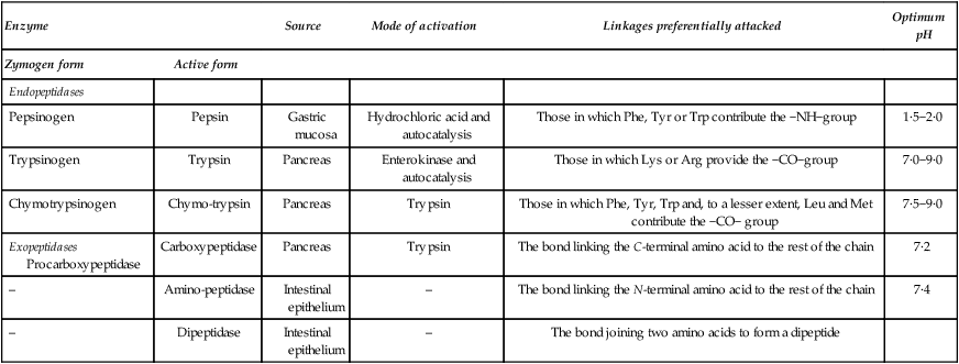
As the food proteins pass along the gastrointestinal tract they undergo a systematic attack, being first subjected to three endopeptidases (pepsin, trypsin and chymotrypsin) which act on proteins and large polypeptides, splitting them at definite points along the chain. The peptides so produced are then subjected to the action of several types of exopeptidase which break off terminal amino acids. A list of the proteolytic enzymes of the gastrointestinal tract is given in Table 19.1.

Table 19.1

The main proteolytic enzymes of the digestive tract

Enzyme Source Mode of activation Linkages preferentially attacked Optimum pH
Zymogen form Active form        
Endopeptidases          
Pepsinogen Pepsin Gastric mucosa Hydrochloric acid and autocatalysis Those in which Phe, Tyr or Trp contribute the −NH−group 1·5−2·0
Trypsinogen Trypsin Pancreas Enterokinase and autocatalysis Those in which Lys or Arg provide the −CO−group 7·0−9·0
Chymotrypsinogen Chymo-trypsin Pancreas Trypsin Those in which Phe, Tyr, Trp and, to a lesser extent, Leu and Met contribute the −CO− group 7·5−9·0
Exopeptidases Procarboxypeptidase Carboxypeptidase Pancreas Trypsin The bond linking the C-terminal amino acid to the rest of the chain 7·2
Amino-peptidase Intestinal epithelium The bond linking the N-terminal amino acid to the rest of the chain 7·4
Dipeptidase Intestinal epithelium The bond joining two amino acids to form a dipeptide  

image

The proteases and particularly the endopeptidases are potentially very dangerous to the organism and must be kept in an inactive state until they have reached the place where they are required at the time that they are required. The endopeptidases and carboxypeptidases, which like trypsin and chymotrypsin are produced in the pancreas, are secreted in precursor or zymogen form which only becomes active after a masking peptide or peptides have been removed.

Although all the peptide bonds between the amino acids in a polypeptide chain are identical they are not all equally susceptible to attack by proteolytic enzymes. These show a distinct preference for certain bonds depending on the nature of the amino acids that participate in the formation of the bonds and, in the case of the exopeptidases, of free amino and/or free carboxyl groupings attached to the α-C atom. Peptides containing proline are particularly resistant to enzymic cleavage and there appear to be specific enzymes for splitting such peptides. Denaturation of proteins by the acid of the stomach, by heating or, as in the case of egg white, by mechanical agitation, causes the highly organized protein molecules to uncoil and expose a larger surface to enzyme attack. However, excessive or prolonged cooking may cause extra linkages to be formed so that the protein becomes less, instead of more, digestible.

Protein digestion starts in the stomach where the acid secreted by the oxyntic cells both assists the denaturation of the proteins and activates the pepsinogen secreted by the peptic cells by removing about 20% of the molecule. Activation is autocatalytic; exposure to the very low pH of the stomach contents also provides optimum conditions for the activity of the enzyme. Pepsin is not very specific but is most active with respect to bonds in which an aromatic amino acid provides the −NH−group. Usually about 10% of the bonds in food proteins are cleaved by pepsin and this produces peptides of molecular weight 600−2000, which are the ‘peptones’ used in preparing bacteriological media.

On passing into the duodenum the chyme from the stomach is mixed with the pancreatic juice which contains trypsin, chymotrypsin and carboxypeptidase in their zymogen forms. Trypsinogen is converted into trypsin either by enterokinase, an enzyme secreted by the duodenal mucosa, or autocatalytically by trypsin itself. In the conversion a hexapeptide Val-(Asp)4-Lys is split off, exposing the active centre of the enzyme which consists of the hydroxyl group of serine in close proximity to two histidine residues. Trypsin is highly specific in its action and only breaks bonds which involve the carbonyl groups of either lysine or arginine.

Chymotrypsinogen, which consists of a single chain containing 246 residues and five disulphide bonds, is activated in several stages. Activation is initiated by trypsin and completed by chymotrypsin itself, the final form consisting of three residual chains held together by disulphide bonds. Chymotrypsin attacks bonds involving the carbonyl groups of the aromatic amino acids, phenylalanine, tyrosine and tryptophan.

Procarboxypeptidase is the inactive precursor of the pancreatic exopeptidase which hydrolyses the peptide bonds joining the amino acid in the C-terminal position to the rest of the chain.

Trypsin is unique among pancreatic enzymes in that it is capable of activating all the pancreatic proenzymes including itself. The pancreas of all mammals contains a potent tryptic inhibitor which protects the gland against autodigestion by small amounts of active trypsin formed within it but which does not prevent proteolysis of food by the fully activated juice.

The oligopeptides formed by the action of the endopeptidases are broken down into their constituent amino acids by the action of the exopeptidases. The carboxypeptidase of the pancreas splits amino acids one by one from the C-terminus so that, by the time they reach the absorbing cells of the small intestine, the dietary proteins have been converted into a mixture of amino acids and small peptides. The mucosal cells which contain both aminopeptidases and dipeptidase take up the small peptides which are then hydrolysed either within the brush border or in the layer immediately beneath it. Thus the final stages of protein digestion, like those of carbohydrates, are intracellular. Under normal circumstances no peptides pass across the mucosa to enter the bloodstream.

Amino acid absorption

Amino acids are rapidly absorbed in the intestine. The intestinal wall is lined with specialized absorptive cells whose primary function is the transport of nutrients from the lumen of the gut into the portal circulation. These cells contain active transport systems for both sugars and amino acids in the brush border membrane.

The transport of most amino acids in the intestine is linked to the transport of Na+ ions in the same direction (symport). The Na+ ions are carried down a concentration gradient on the same carrier as the amino acids which are carried against a concentration gradient. The inward Na+ gradient is maintained by the action of a Na+/K+-ATPase which pumps the Na+ ions out of the cell in exchange for K+ ions.

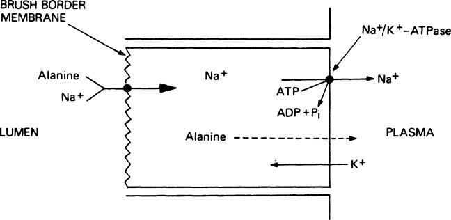
Thus the energy for the active transport of amino acids is derived indirectly from the hydrolysis of ATP (Figure 19.1). After uptake into the absorptive cells by this method, the amino acids pass into the portal circulation by a passive transport process. The brush border membrane contains a number of different transport systems for amino acids, which have overlapping specificity. It is probable that similar systems are responsible for the uptake of amino acids into other tissues such as kidney and liver. As a result of the operation of such transport systems, the total free amino acid concentration in the plasma is kept at between 2 and 4 mM but in the tissues it is between 15 and 30 mM. Amino acids derived exogenously from the food are mixed with amino acids derived endogenously from the tissues to form a metabolic pool. The essential amino acids (see below) are taken up by the tissues with great avidity and the non-essential amino acids, notably glycine, alanine, glutamic acid and glutamine, account for 80% of the total free amino acid nitrogen.

The absorption of intact protein

Occasionally large polypeptides and even complete proteins are absorbed. These cannot be used for protein synthesis and may lead to immunological sensitization and allergic reactions. Absorption of native protein is, however, a normal process in certain newborn animals. In Man passive immunity is conferred on the newborn infant by placental transfer of maternal antibodies. In some other species including the cow, horse, goat, sheep and pig this does not occur and, instead, antibodies are supplied in the colostrum, the secretion of the mammary glands which is produced prior to the start of lactation proper. The colostrum proteins are protected against digestion by the presence in colostrum of a trypsin inhibitor and also by the failure of the neonate to secrete hydrochloric acid. In the first few hours the proteins are absorbed in large quantities by pinocytosis but after about 36 hours the intestine loses its ability to absorb intact protein.

Essential amino acids

Although plants and many microorganisms are able to synthesize all the amino acids they require from simple carbon compounds and non-specific sources of nitrogen such as ammonia, higher animals are unable to do this and must obtain some of the amino acids from the protein in the diet. An essential amino acid is one that an animal is either unable to synthesize for itself or which it cannot synthesize at a sufficient rate to meet the needs for metabolism and growth. Different species vary to some extent in their essential amino acids. Humans require at least eight and probably ten. They are valine, leucine, isoleucine, lysine, methionine, phenylalanine, threonine and tryptophan with arginine and histidine having a doubtful status.* The latter are ‘relatively indispensable’ in that they can be synthesized in the body but their rate of synthesis may be too slow fully to supply the needs of the growing child. On a similar basis glycine is an essential amino acid for rapid growth of feathers in young birds such as the chick. Young rats require the same ten amino acids as are needed by children.

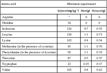
The quantities of the various essential amino acids required daily in the diets of individual humans were determined by Rose who fed young adult volunteers on diets containing adequate amounts of purified carbohydrate, fats, minerals and vitamins but provided mixtures of amino acids in the place of protein. The mixtures contained all the amino acids except the one under investigation which was given separately and, by slight alterations in the level of intake, the subject could be made to go reproducibly from positive to negative balance and back again. This indicated the daily requirement of the individual for this amino acid. The experiment was repeated for each essential amino acid in turn thus giving a picture of the overall needs of that person. Particular individuals were found to have well-defined requirements although there was considerable variation from one person to another. Average results for men, women and infants are given in Table 19.2. As mentioned in Chapter 10 the ‘safe’ level of a given amino acid is taken as being considerably higher than the minimum requirement.

Table 19.2

Estimated daily requirements for the essential amino acids

Amino acid Minimum requirement
  Infants(mg kg−1) Men(g) Women(g)
Arginine * 0 0
Histidine 34 0 0
Isoleucine 126 0·7 0·55
Leucine 150 1·1 0·73
Lysine 103 0·8 0·54
Methionine (in the presence of cysteine) 45 1·1 0·70
Phenylalanine (in the presence of tyrosine) 90 1·1 0·70
Threonine 87 0·5 0·37
Tryptophan 22 0·25 0·17
Valine 105 0·8 0·62

image

*No figure given

The requirements for the essential amino acids are further complicated by the finding that two non-essential amino acids can only be synthesized in the body if two of the essential amino acids are present in sufficient amounts. Tyrosine is formed directly from phenylalanine so that the requirement for phenylalanine is less when tyrosine is present than when it is absent from the diet. Similarly the sulphur that is required for the synthesis of cysteine can only be obtained from methionine so that the dietary requirements for the sulphur-containing amino acids should be considered together. If cysteine is present in ample amounts the requirement for methionine will be minimal, but if cysteine is in short supply more methionine is needed.

It should perhaps be pointed out that the ‘non-essential’ amino acids are just as important in metabolism as the ‘essential’ amino acids, the distinction being the need for an external supply of the latter. If protein is to be synthesized, all its constituents must be simultaneously available and experiments have shown that if a missing essential amino acid is fed an hour or so after the others it is inefficiently utilized.

In most instances it is the α-oxo acid corresponding to the essential amino acid that the body is unable to synthesize and, if this oxo acid is supplied it can be quite readily converted into the corresponding amino acid. Exceptions to this rule are lysine and threonine which have to be supplied in the amino acid form.

Amino acid metabolism

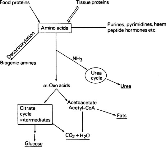
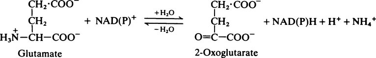
A summary of amino acid metabolism is given in Figure 19.2. Amino acids are used for protein synthesis and as N and C donors for the synthesis of other types of macromolecule, e.g. the nucleic acids as well as numerous small molecular compounds. After deamination, i.e. removal of the amino group, the carbon skeleton may be used for the formation of glucose or even fats or it may be oxidized to CO2 and water with the production of metabolic energy. Decarboxylation, i.e. removal of the carboxyl group of certain of the amino acids, leads to the production of biogenic amines such as histamine, serotonin and γ-aminobutyrate.

Tissue interrelationships in amino acid metabolism

While dietary carbohydrate in excess of immediate requirements may be stored in the form of glycogen, and lipid may be stored as triglyceride, there is no analogous short-term storage of amino acids. Amino acids in excess of requirements are immediately degraded. In fact, it has been shown that after eating a balanced meal, degradation of excess amino acids precedes the metabolism of carbohydrates and fats.

The breakdown of amino acids involves the liberation of the α-amino group in the form of ammonia. Ammonia is extremely toxic, especially, for reasons which are still not fully understood, to the brain and one of the major functions of the mammalian liver is to detoxify ammonia by converting it to urea (CO(NH2)2); this is non-toxic, highly water-soluble and readily excreted via the kidneys. Different mechanisms of ammonia detoxification occur in fish where ammonia is excreted directly via the gills, and in birds and reptiles where ammonia is converted to uric acid. Uric acid is insoluble in water and is thus suitable for excretion under conditions where the water supply may be limited.

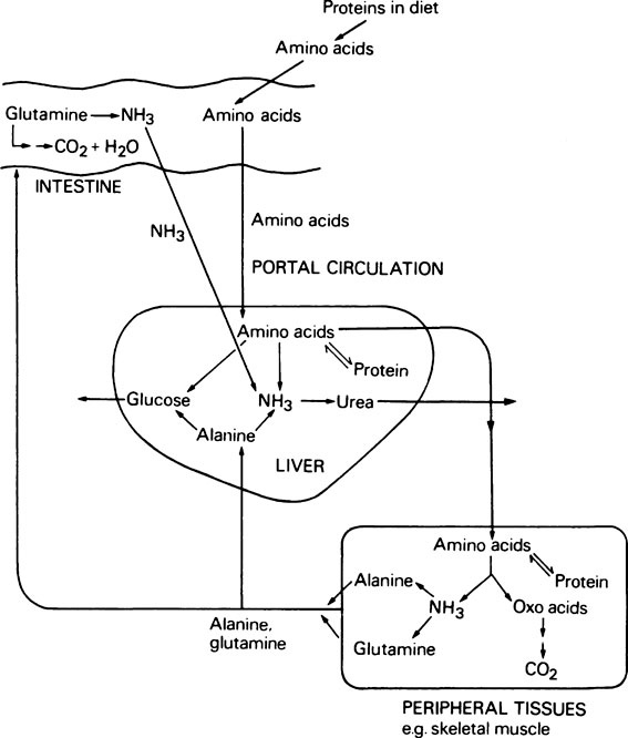
The metabolism of amino acids proceeds by pathways which are common to most tissues, but the pathway for the conversion of ammonia to urea occurs only in the liver. During the degradation of amino acids in peripheral tissues such as skeletal muscle, the ammonia formed is not released directly into the bloodstream. Instead it is used to form the amino acids alanine and glutamine from pyruvate and glutamate which are readily available, and it is these amino acids that are then released. The alanine produced by the tissues is taken up by the liver and converted to urea and glucose. Although some glutamine is metabolized by the liver, the major site of glutamine metabolism is the intestine where it is used as a major respiratory fuel. The ammonia produced by glutamine metabolism in the gut returns immediately via the portal circulation to the liver, where it is detoxified. These tissue interrelationships in amino acid degradation are illustrated in Figure 19.3.

Pathways of amino acid catabolism

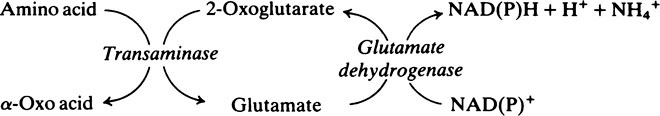
Transamination and deamination

The initial step in the degradation of many amino acids is a transamination reaction whereby the α-amino group of the amino acid is transferred to α-oxoglutarate with the formation of glutamate and the α-oxo acid corresponding to the amino acid in question. Transamination reactions are catalysed by a group of enzymes called transaminases or aminotransferases. The most important of these are glutamate−oxaloacetate transaminase (GOT) and glutamate−pyruvate transaminase (GPT); the reactions catalysed by these enzymes are shown below.

< ?xml:namespace prefix = "mml" />CH3·CO·CH2·COO+ATP+CoASHThiokinaseCH3·CO·CH2·COSCoA+AMP+PPi

image

CH3·CO·CH2·COO+ATP+CoASHThiokinaseCH3·CO·CH2·COSCoA+AMP+PPi

image

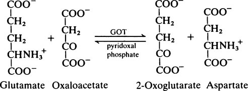
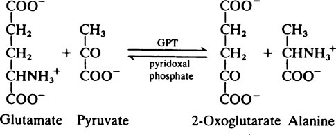
Transaminase reactions are freely reversible so that they function in both the synthesis and breakdown of amino acids. All transaminases require pyridoxal phosphate, a derivative of vitamin B6 (page 165), as a cofactor which transfers the α-amino group from an amino acid to a keto acid. In general, transaminases have a high Km value for the appropriate amino acid but a much lower Km for 2-oxoglutarate.

The glutamate formed by the transamination of amino acids with 2-oxoglutarate then undergoes oxidative deamination via the glutamate dehydrogenase reaction:

CH3·CO·CH2·COO+ATP+CoASHThiokinaseCH3·CO·CH2·COSCoA+AMP+PPi

image

Glutamate dehydrogenase is a mitochondrial enzyme which can use either NAD+ or NADP+ as the hydrogen acceptor.

Although the equilibrium of this reaction is very much in favour of glutamate formation, in the cell the rapid removal of the 2-oxoglutarate and NAD(P)H allows the enzyme to function efficiently in the direction of glutamate deamination. Liver glutamate dehydrogenase is a very active enzyme, and the reaction is not rate-limiting for amino acid deamination.

Glutamate dehydrogenase is responsible not only for the deamination of glutamate itself but also indirectly for the deamination of many other amino acids. For example, when alanine is transaminated with 2-oxoglutarate, pyruvate and glutamate are produced. The glutamate is then deaminated via the glutamate dehydrogenase reaction and 2-oxoglutarate is regenerated and is available to transaminate with another molecule of amino acid. The net result of the two reactions is the deamination of one molecule of the amino acid with the production of one molecule of its corresponding α-oxo acid and one molecule of ammonia and the reduction of one molecule of NAD(P)+.

CH3·CO·CH2·COO+ATP+CoASHThiokinaseCH3·CO·CH2·COSCoA+AMP+PPi

image

Not all amino acids are deaminated in this way. The amino groups of glutamine and asparagine are directly hydrolysed by the enzymes glutaminase and asparaginase with the production of ammonia. The hydroxyamino acids serine and threonine are acted upon by the enzymes serine dehydratase and threonine dehydratase respectively, again with the direct production of ammonia, while proline, arginine and histidine are metabolized to form glutamate and the amino group is then removed by glutamate dehydrogenase.

Stay updated, free dental videos. Join our Telegram channel

Dec 10, 2015 | Posted by in General Dentistry | Comments Off on The metabolism of proteins and amino acids

VIDEdental - Online dental courses

Get VIDEdental app for watching clinical videos