History of the Procedure
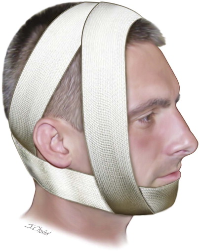
The concept of immobilization for the treatment of skeletal fractures dates back to the days of Ancient Greece as documented by Hippocrates. Fixation methods have evolved over time through the use of the Barton bandage ( Figure 59-1 ) and Gunning splints. Guglielmo Salicetti and Gilmer are thought to be among the first to make use of intermaxillary wires for the treatment of mandible fractures. Although the advent of rigid bone plates has dramatically changed the way mandibular and other facial fractures are treated, the use of intermaxillary fixation remains a time-tested and often invaluable adjunct in the treatment of facial trauma.
History of the Procedure
The concept of immobilization for the treatment of skeletal fractures dates back to the days of Ancient Greece as documented by Hippocrates. Fixation methods have evolved over time through the use of the Barton bandage ( Figure 59-1 ) and Gunning splints. Guglielmo Salicetti and Gilmer are thought to be among the first to make use of intermaxillary wires for the treatment of mandible fractures. Although the advent of rigid bone plates has dramatically changed the way mandibular and other facial fractures are treated, the use of intermaxillary fixation remains a time-tested and often invaluable adjunct in the treatment of facial trauma.

Indications for the Use of the Procedure
Intermaxillary fixation (IMF) is indicated for any procedure in which intact dentition or jaw can be used to assist in the reduction or realignment of the opposing dentition and arch. It can be employed either as an aid to open reduction of mandible fractures or as definitive treatment for mandibular fractures not amenable to open reduction. These situations include minimally displaced favorable fractures, grossly comminuted fractures, pediatric mandibular fractures, and intracapsular condylar fractures. Although dentate jaws are ideal, IMF can also be accomplished for the edentulous arch through the use of Gunning splints or existing dentures. IMF is also indicated for the reapproximation and reduction of panfacial fractures for which dentition can be used as a template for definitive treatment. In addition, IMF is a useful treatment intermediary for orthognathic and other reconstructive procedures ( Figure 59-2 ).
Limitations and Contraindications
As with any other surgical procedure, intermaxillary fixation is not without its limitations. Prolonged immobilization has been associated with trismus and decreased range of motion in the postfixation period with the need for extensive physical therapy and temporomandibular joint (TMJ) exercises. Many patients find it difficult to maintain good oral hygiene in the presence of wires and arch bars in the oral cavity. Ultimately, a small subset of patients will be unable or unwilling to tolerate intermaxillary fixation for any period of time. Those patients include the elderly, obtunded patients, developmentally delayed patients, and alcoholics. Patients prone to nausea and vomiting are at risk for aspiration and may be best treated with other methods of fixation. Although edentulous patients can be treated with IMF through the use of splints, most surgeons prefer to treat edentulous fractures with rigid plates, forgoing the laborious and time-consuming task of either modifying existing dentures or fabricating Gunning splints.
Intracapsular condylar fractures should undergo only limited durations of IMF for pain relief, assuming the occlusion is stable and reproducible. TMJ ankylosis, though seldom seen, is a feared complication of prolonged intermaxillary fixation. Airway concern may be a relative contraindication, especially in the obtunded trauma patient. A tracheostomy should be considered to provide a secure airway in the multitrauma patient for whom an extended period of intubation is anticipated.
Injuries in the pediatric population deserve special mention. IMF times in pediatric mandibular fractures should be shorter given their excellent healing and bone formation capacities. This is especially true for condylar fractures in children due to their propensity to grow heterotopic bone and undergo ankylosis.
Technique: Application of Erich Arch Bars
Step 1:
Intubation and Preparation
Erich arch bars should be trimmed to length, typically from at least the first molar to the first molar on either side. Longer spans may be required to stabilize fracture segments. Either 24- or 26-gauge wire should also be cut for interdental and interarch fixation. Once the hardware has been prepared, intermaxillary fixation can be completed using general anesthesia, IV sedation, or local anesthesia alone. Along with the soft tissue and dentition to be wired, the fracture sites should be anesthetized using local anesthesia with a vasoconstrictor, paying close attention to recommended maximum dosages.
Step 2:
Application of Arch Bar
Once adequately anesthetized, the arch bar can be fixed to each tooth using pretrimmed 24-gauge wire. The wire can be passed from labial to lingual on one side of the tooth and passed back from lingual to labial on the opposing surface. The wire should rest superior to the bar on one side and inferior to the bar on the other, effectively creating a loop, which can be tightened against the arch bar. This is repeated for all teeth within the span of the arch bar on both the mandible and maxilla. At this point, any teeth scheduled for removal should be extracted. Judicious removal of teeth should be undertaken, because the literature fails to show a significant difference in infection rates when teeth in the line of fracture are extracted or retained ( Figure 59-3, A ).
Stay updated, free dental videos. Join our Telegram channel
VIDEdental - Online dental courses


