Signal Molecule-Calcium Phosphate Composites: Novel Approaches to Controlling Cellular and/or Biological Reactions and Functions
(6.1)
where I is the ionic activity product of the solution with respect to a CaP, Ks is the solubility product of the CaP, and ν is number of ionic activity in the product (e.g., ν = 5 and 9 for (Ca2+)3(PO43−)2 and (Ca2+)5(PO43−)3(OH−), respectively). Physiologically supersaturated CaP solutions such as body fluid, saliva, Kokubo’s simulated body fluid (SBF), and Hank’s solution are all supersaturated with respect to Ap and octacalcium phosphate (OCP) and nearly saturated with respect to amorphous calcium phosphate (ACP) in the pH range of 7.3–7.4 at 25 °C: for example, the S values of SBF are 12–19, 0.6–2.5, and −0.4 for Ap, OCP, and ACP, respectively, at pH 7.4 and 25 °C. Supersaturated CaP solutions contain prenucleation CaP clusters under physiological pH condition [1–5], which accounts for S being as high as 12–19 with respect to Ap. Owing to such a high value of S, Ap can be formed by both direct condensation of ions (classical route) and formation of precursors such as cluster aggregates or ACP followed by transformation into Ap (multistep route) [6].
Molecules present in a supersaturated CaP solution exhibit a variety of interactions with CaP crystals, precursors, and/or clusters in the solution depending on solution chemistry, pH, and temperature, which is yet to be fully understood. A protein molecule, a CaP crystal, precursors of the CaP crystal, and clusters can have a positive or a negative surface charge depending on solution chemistry and pH. It is when the molecule and CaP have opposite electrical charges that interaction can occur [7]. Thus, for example, the efficiency of coprecipitation of basic proteins largely differs between different supersaturated CaP solutions [8–10]. Temperature is another factor that influences the efficiency of coprecipitation owing to the temperature dependence of solubility and stability of CaPs [11]. Two different mechanisms have been proposed for the process of coprecipitation: one is molecular adsorption followed by crystal nucleation [12–14], and the other is molecular incorporation into ACP followed by amorphous-crystalline transformation [15]. The former is proposed by analogy with the fact that many noncollagenous acidic matrix proteins in bone promote Ap formation when they are immobilized on a substrate while they inhibit Ap formation when they are unimmobilized. Thus, once molecules are adsorbed on precipitated CaP, the adsorbed molecules act as the secondary nucleation sites of CaP crystals in a supersaturated CaP solution. In the latter mechanism, molecules, calcium ions, phosphate ions, and clusters are aggregated into an amorphous phase. As the molecule-containing ACP transforms to a polycrystalline phase, the molecules are captured on the surface or interstices of crystals. In both mechanisms, the final product is a nanocomposite of molecules and CaPs where the molecules are dispersed in the tens-to-hundred-nanometer-scale distances within the CaP matrix (Fig. 6.1) [12, 16].
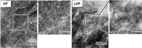
Fig. 6.1
Transmission immunoelectron microscopic photographs for an Ap layer (CP) and a laminin-Ap composite layer (LCP). Black dots in LCP correspond to laminin molecules (Reprinted from Oyane et al. [16], Copyright 2006, with permission from Elsevier)
Molecules thus immobilized so far include fibronectin [12], vitronectin [12], albumin [17, 18], collagen [19], amelogenin [20], laminin [21], BMP-2 [22], FGF-2 [10, 15], antibiotics [23, 24], l-ascorbic acid phosphate magnesium salt n-hydrate (AsMg) [25–27], DNA [28], α-amylase [29], and immunogenic molecules [30].
The molecule present in a supersaturated CaP solution controls the phase, size, and crystallinity of CaP precipitated. Albumin facilitated Ap crystallization in a supersaturated CaP solution where only OCP is formed in the absence of albumin [17]. Proteins that have particular peptide motifs present in dentin matrix protein 1 facilitated the transformation of ACP into Ap [31]. However, many molecules decrease the size and crystallinity of Ap or inhibit Ap crystallization [21, 24, 25, 32].
Supersaturated CaP solutions used for fabricating signal molecule-CaP composites are classified into (1) physiological saline, (2) SBF-based CaP solutions, (3) inhibitor-free CaP solutions, and (4) infusion fluid-based CaP solutions. Examples of physiological saline include Hanks’ balanced salt solution, Dulbecco’s phosphate-buffered saline, and phosphate-buffered saline (PBS) [14, 33–35]. SBF is a supersaturated CaP solution that has ion concentrations approximately equal to those of human blood plasma. 1.5 times concentrated SBF is prepared at pH 7.25. SBF with doubled Ca and P concentrations is prepared at pH 6.8 [36]. Bubbling carbon dioxide gas at a pressure of 0.1 MPa allows preparation of SBF concentrated as high as seven times [37]. Once supply of carbon dioxide gas is stopped, the gas is released out of the solution, leading to an increase in pH, thus increasing supersaturation and enhancing formation of CaP [37–39]. SBF and concentrated SBF are used for fabricating signal molecule-CaP composites or base coating of signal molecule-CaP composites [17, 40]. Physiological saline and SBF-based CaP solutions contain not only calcium and phosphate ions but also magnesium and carbonate ions that inhibit CaP formation. Removal of these inhibitor ions (inhibitor-free CaP solutions) accelerates formation of signal molecule-CaP composites [17, 21, 28, 41]. Physiological saline, and SBF-based and inhibitor-free CaP solutions contain organic buffering agents such as tris(hydroxymethyl)aminomethane (TRIS) and 4-(2-hydroxyethyl)-1-piperazineethanesulfonic acid (HEPES) except for those buffered with carbon dioxide gas. Besides buffering agents, these solutions do not necessarily guarantee biological safety for clinical use, owing to possible contamination by harmful impurities, microorganisms, viruses, and pyrogens. Infusion fluid-based CaP solutions have the advantage of biological safety over other classes of supersaturated CaP solutions. Infusion fluids are sterile, contain only trace amounts of pyrogens, and are approved officially for clinical use of injection. Aseptically mixing calcium-containing, phosphate-containing, and pH-adjusting infusion fluids allows the preparation of supersaturated CaP solutions that have a high level of biological safety [11, 19, 26, 42].
6.2.2 Material Properties of Signal Molecule-CaP Composite Layers
The CaP phase in signal molecule-CaP composite layers includes low-crystalline Ap [21, 24, 43], ACP [8], OCP, and/or an OCP-like phase [18, 20, 43, 44]. The thickness of the composite layer is from several hundred nanometer to some tens microns. The morphology and crystallinity of the matrix phase change with process parameters including solution chemistry and fabrication temperature. The Mg ion and molecule in the solution decrease the surface roughness of the layer through their inhibitory effect on the crystal growth of Ap, which results in a decrease in the crystal size of the formed Ap [45].
Information regarding the mechanical properties of signal molecule-CaP composite layers is available for some composite layers. The shear strength of the laminin-Ap composite layer depends on the moisture condition: the shear strength of the laminin-Ap composite layer is higher than that of the Ap layer under wet condition, while that of the former is lower than that of the latter under dry condition [46]. The adhesion strength values for 3- and 10-μm-thick laminin-Ap composite layers are 7 and 2 MPa, respectively, under dry condition, which are both lower than those for 3- and 10-μm-thick Ap layers, respectively [46]. Coatings with a Ca/P molar ratio of 1.30 and thicknesses of 15–23 μm prepared in supersaturated CaP solution in the presence of bovine serum albumin (BSA) had significantly higher critical loads in the scratch test than those with a Ca/P molar ratio of 1.33 and a thickness of 33 μm prepared in the absence of BSA [47]. However, the hardness of these coatings was not significantly influenced by the presence of BSA. A BMP-2-CaP composite layer had significantly higher critical loads (2.5 ± 0.37N) in the scratch test than the CaP layer (1.8 ± 0.08N) prepared in the absence of BMP-2 [44].
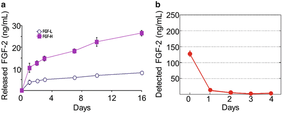
Immobilization enables the localization of signal molecules at specific body sites and their release in a sustained manner. On the contrary, free molecules cannot be retained at a local site for a long time because they rapidly diffuse from the site [48, 49]. Albumin [17], cytochrome c [8], and FGF-2 [50] immobilized in the signal molecule-CaP composite layers are released from the composite layers in physiological sodium chloride solution and cell-culture media (Fig. 6.2a). The release was sustained for 10–21 days, while these molecules adsorbed to the same substrate were released much faster and in some cases completely within 24 h. Collagen and fibronectin were immobilized firmly on an Ap ceramic and continued to be released into a physiological saline solution for at least 3 days [19, 51]. The DNA-CaP composite coating exhibited an initial burst of release during the first 8 h and a sustained release for a few days after exposure to a cell-culture medium [28, 52]. An ascorbate-Ap composite coating resulted in slower ascorbate release in physiological saline solution than in the case of adsorbed ascorbate [53].
Fig. 6.2
(a) Cumulative release of FGF-2 from FGF-2-Ap composite layers containing FGF-2 at a low dose (FGF-L) and a high dose (FGF-H) in DMEM at 37 °C (Reprinted from Tsurushima et al. [50], Copyright 2010, with permission from Elsevier). (b) Decomposition of unimmobilized FGF-2 in DMEM at 37 °C
The biological activity of immobilized molecules is either decreased, preserved, or increased compared with that of unimmobilized ones. Biological activity may be decreased or lost when the molecular chemistry or configuration is changed by immobilization. This typically occurs when the chemical bonding of molecules to a substrate is too strong or the immobilizing reaction is degradative. Biological activity may be preserved when the molecular chemistry and configuration are unchanged by immobilization. The activity can be preserved even for a longer period than unimmobilized molecules since many biologically active and unimmobilized molecules have short lifetimes and lose their activity with time by natural denaturation or decomposition under physiological condition [54]. For example, in Dulbecco’s Modified Eagle Medium (DMEM), the FGF-2-Ap composite layer preserved and released FGF-2 that is detectable with enzyme-linked immunosorbent assay (ELISA) even after 14 days [50, 55], while unimmobilized FGF-2 added directly to DMEM decomposes over 95 % within 48 h (Fig. 6.2b). The biological activity of immobilized molecules may be increased compared with unimmobilized molecules owing to increases in the molecular stability and local concentration, multivalency, and inhibition of cellular internalization of the molecules [54, 56].
The biological activity of signal molecule-CaP composites is characterized by bioassay. In particular, in vitro bioassay that reflects in vivo biological activity is useful and can be used for quality control of signal molecule-CaP composites. In vitro bioassay includes evaluation of the mitogenic activity of NIH3T3 and BHK-21 cells cultured with FGF-2-CaP composite layers [10, 27, 50], evaluation of the alkaline phosphatase (ALP) activity of rat bone marrow stromal cells cultured on BMP-2-CaP composite layers [22], and evaluation of cytokine secretion by THP-1 cells cultured with immunogenic molecules-CaP composites [30, 57].
6.3 Applications to Drug Delivery for Tissue Regeneration
Tissue loss or failure is one of the most frequent, devastating, and costly problems in human health care. Tissue engineering, which applies the principles of biology, engineering, and materials science, has emerged as potential means of growing new tissues and organs [58–60]. Signal molecules that regulate a variety of cellular processes, e.g., cell adhesion, proliferation, differentiation, and maturation, are crucial for tissue engineering or regenerative medicine to regulate cellular functions [58]. Immobilization of signal molecules with CaP on a substrate is a promising process to achieve a sustained regulation activity of the signal molecules [43]. To date, signal molecules have been integrated into CaP coatings for the purpose of promoting bone regeneration and soft tissue regeneration.
6.3.1 Bone Regeneration
Bone defects have been treated by surgery, using bone grafts such as autologous bone, allografts, or synthetic bone graft materials [61–63]. The major drawbacks of autologous bone are their limited supply and surgical side effects. Allografts have concerns related to host response and disease transmission and can be associated with infection and inflammation [64, 65]. Synthetic bone graft materials, typically CaPs, have no or clinically insufficient osteoinductive property. Tissue-engineered bone and biomaterials loaded with signal molecules are important alternatives. As one of the latter class, signal molecules such as BMP-2, FGF-2, ascorbate, collagen, fibronectin, amelogenin, and poly(l-lysine) are coprecipitated with CaPs to be immobilized on biomaterials to increase bone cell attachment, proliferation, and tissue regeneration.
BMP-2, a protein that induces the formation of bone and cartilage, is widely used for bone regeneration. BMP-2 in the BMP-2-CaP composite layers retains its bioactivity, e.g., it stimulated ALP activity of rat bone marrow stromal cells when the cells were seeded directly on these layers for 8 days [22, 44, 66, 67]. BMP-2-CaP composite layers are highly biocompatible, osteoconductive, and osteoinductive, and they promote ectopic bone formation [22, 44, 66, 67].
FGF-2 is one of several well-characterized growth factors; it is highly expressed in developing tissues, stimulates vascular endothelial cell proliferation and neovascularization, and regulates cell growth, differentiation, and migration [68–70]. FGF-2 at an appropriate dose can promote bone regeneration. FGF-2-CaP composite layers promoted the proliferation of osteoblastic MC3T3 and MG63 cells [34, 50]. These enhanced bone formation at an optimized FGF-2 dose compared with layers without FGF-2 in full-thickness skull defects in rats [50].
l-Ascorbic acid phosphate magnesium salt n-hydrate (AsMg)-Ap composite layers markedly enhanced osteoblastic MC3T3-E1 cell proliferation and differentiation in vitro [27]. Collagen-Ap composite layers obviously enhanced the attachment, proliferation, and differentiation of Saos-2 osteoblast-like cells [71–74]. Collagen-fibronectin-CaP composite layers promoted osteoblastic cell proliferation and differentiation and new bone formation in full-thickness skull defects in rabbits [19, 51]. Amelogenin-Ap composite layers increased type I collagen gene expression level, ALP activity, and osteocalcin content of human embryonic palatal mesenchymal preosteoblast cells [20, 75]. BSA-CaP composite layers showed high biocompatibility and osteoconductivity [76, 77]. Zinc/silicate-Ap composite layers enhanced the proliferation and differentiation of osteoblastic MC3T3-E1 cells [78, 79]. Poly(l-lysine)-, poly(l-glutamic acid)-Ap composite layers promoted initial osteoblast adhesion, spreading, and proliferation, and were critical to forming a stable tissue implant interface [80].
6.3.2 Soft Tissue Regeneration
Signal molecules are used as stimulators of soft tissue regeneration. Signal molecule-CaP composites are used for promoting soft tissue formation around percutaneous implants, accelerating vascularization in cerebral infarction lesions, promoting epithelial cell adhesion, and preventing epidermal downgrowth, among others.
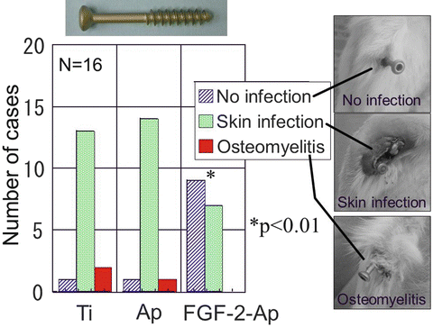
Accelerated cell adhesion and tissue regeneration at the interface between the external fixation pin, a percutaneous implant, and soft tissues are required for decreasing infection around the percutaneous device. Several signal molecules were immobilized with Ap on titanium (Ti) external fixation pins to accelerate cell proliferation, differentiation, and tissue regeneration around the pin. Ti rods, which correspond to the stem part of the Ti external fixation pin, coated with AsMg-FGF-2-Ap composite layers markedly enhanced proliferation and procollagen type І gene expression of fibroblastic NIH3T3 cells [27]. Ti rods coated with zinc/silicate-Ap composite layers accelerated proliferation of fibroblastic NIH3T3 and osteoblastic MC3T3-E1 cells and differentiation of MC3T3-E1 cells in vitro [78, 79]. Ti screws coated with FGF-2-Ap composite layers exhibited a significantly higher bone-screw interface strength and a lower pin-tract infection rate than uncoated titanium screws after percutaneous implantation into the proximal tibial metaphyses in rabbits for 4 weeks (Fig. 6.3) [81].
Fig. 6.3
Pin-tract infection around percutaneously implanted uncoated Ti screws (upper left, Ti) and those coated with Ap (Ap) and FGF-2-Ap composite layers (FGF-2-Ap). Infection rate for Ti screws coated with FGF-2-Ap composite layers was 44 % (7/16), while that for uncoated and Ap-coated Ti screws was 94 % (15/16), which is statistically significant (p < 0.01) [81]
Ti screws coated with the FGF-2-Ap composite layers showed a significantly higher wound healing rate than those coated with an Ap layer [82]. Ti screws coated with an AsMg-FGF-2-Ap composite layer significantly reduced the infection rate compared with those coated with an Ap layer after percutaneous implantation of the screws in both proximal tibial metaphyses in rabbits for 4 weeks [43]. It is suggested that the enhanced wound healing associated with the formation of Sharpey’s fiberlike tissue triggered by FGF-2 leads to the reduction in pin-tract infection rate [82].
Besides Ti, stainless steel was coated with AsMg-Ap composite layers using an interfacial layer of mesoporous bioactive glass existing between the metal and composite layers. The stainless steel coated with AsMg-Ap composite layers enhanced the proliferation of fibroblastic NIH3T3 cells in vitro [26]. Titanium or ethylene-vinyl alcohol copolymer (EVOH) coated with laminin-Ap composite layers showed greater epithelial cell adhesion than that uncoated and coated with Ap [21, 41]. The laminin-Ap composite layer is promising for preventing bacterial invasion through the interface around percutaneous implants such as dental implants, as adhesion of epithelial cells to surfaces of teeth or dental implants prevents bacterial invasion [83, 84]. EVOH coated with laminin-Ap composite layers had higher adhesion strength to the skin of rat than EVOH coated with Ap when they were percutaneously implanted [85]. However, there was no significant difference in epidermal downgrowth (which likely leads to bacterial infection through the interfacial space) between EVOH coated with laminin-Ap composite layers and EVOH coated with Ap.
Although FGF-2 is promising for treating brain infarction, intravenously administered FGF-2 has a short circulation half-life (0.5–3 min) and is sequestered rapidly in several organs [86]. An FGF-2-Ap composite layer was applied to prevent the progress of brain infarction in a rat cerebral ischemia model for progressive cerebrovascular disorder, called moyamoya disease [55]. The treatment of moyamoya disease includes the introduction of blood vessels to the brain from the soft tissue outside the skull through an opening or burr holes created in the skull. Burr hole plugs coated with FGF-2-Ap composite layers were implanted in the animal model. The plugs coated with an FGF-2-Ap composite layer showed an improved effectiveness of preventing the progress of brain infarction. The coated plugs yielded significantly smaller areas of brain ischemia and better capillary density than uncoated plugs and uncoated pugs plus direct FGF-2 administration [55].
6.4 Applications to Gene Delivery
6.4.1 Gene Delivery: An Introduction
Gene delivery is a fundamental technique in molecular biology for manipulating cellular characteristics, functions, and behavior by introducing foreign genes (DNA, RNA) into cells with the aid of viral or nonviral vectors. Table 6.1 shows major gene delivery systems. Physical gene delivery systems such as electroporation [87], microinjection [88], and gene gun [89] are excluded in this chapter.
Viral vectors such as adenoviruses, retroviruses, and lentiviruses are the most efficient. Viral vectors with a few exceptions (e.g., adenovirus) generally produce stable transgene expression almost permanently by introducing a foreign gene into the host cell’s genome [90, 91]. A viral gene delivery system is a well-established method to manipulate cells and has long been studied for gene therapy applications. However, the viral systems have drawbacks such as toxicity (immunogenicity and oncogenicity), transgene overexpression, and difficulty in handling and large-scale production of recombinant viruses [90, 91].
Nonviral vectors (typically a plasmid including therapeutic genes) are safer and easier to use than viral vectors. Generally, nonviral vectors induce transgene expression only transiently (shorter than the cell lifespan) without affecting the host cell’s genome. To enhance delivery efficiency, nonviral vectors are usually used in complex with transfection reagents such as lipids (of either liposomal or nonliposomal type), cationic polymers, and CaPs [92–94]. Nonviral gene delivery (also called transfection) systems have gained increased attention in tissue engineering applications as a tool to control cell behavior including multiplication and differentiation.
Among various nonviral gene delivery systems developed hitherto, CaP-based systems have the advantages of safety, good biocompatibility, and a relatively simple and cost-effective production process. In the following sections, CaP-based gene delivery systems will be described with emphasis on a new system using DNA-CaP composite layers [95].
6.4.2 Gene Delivery Systems Using CaP Composite Layers
A conventional CaP-based gene delivery system [94] is generally mediated by nano-/microparticles of DNA-CaP composites. The DNA-CaP composite particles can be fabricated by mixing a phosphate ion solution and a calcium ion solution that is supplemented with DNA under neutral pH in a controlled manner. Thus precipitated DNA-CaP composite particles are either added to a culture medium bathing cells (Fig. 6.4 (left)) or injected into the body for the delivery of therapeutic genes into cells. The critical disadvantages of CaP-based gene delivery systems are their insufficient efficiency and poor reproducibility compared with other nonviral systems. Various approaches have been proposed to increase the efficiency of CaP-based gene delivery systems. For example, inhibitors of CaP crystal growth, i.e., block copolymers [96] or specific ions such as magnesium and carbonate [97, 98
Only gold members can continue reading. Log In or Register to continue
Nov 10, 2015 | Posted by mrzezo in General Dentistry | Comments Off on Signal Molecule-Calcium Phosphate Composites: Novel Approaches to Controlling Cellular and/or Biological Reactions and Functions