Introduction
Patient photos and silhouettes are commonly used in clinical evaluations and orthodontic research to evaluate profile esthetics. The purpose of this study was to determine whether the use of photos or silhouettes is a more appropriate method of evaluating African American profile esthetics and whether there are different profile esthetic preferences among clinicians when using photos compared with silhouettes.
Methods
Pretreatment records of 20 adolescent African American patients were selected (10 male, 10 female) from the orthodontic clinic at the Albert Einstein Medical Center in Philadelphia. Each patient’s profile photo was digitally changed with imaging software (Dolphin Imaging and Management Solutions, Chatsworth, Calif) to fabricate a series of 7 photos and 7 silhouettes with lip positions at uniform distances relative to Ricketts’ E-line standard. Fifteen raters consisting of orthodontic faculty and residents were asked to select the most esthetically pleasing profile from each patient’s photo series and silhouette series.
Results
More rater preferences for the photographs (86%) were within the acceptable esthetic range (within 2 mm of the E-line in either direction) than were their preferences for silhouettes (66%) ( P <0.001). Flatter profiles with less lip projection than the esthetic norm were more often preferred in the silhouettes than in the photos. Thirty-one percent of the silhouettes preferred by the raters were flatter than the norm compared with 9% of the photos ( P = 0.003). Fuller profiles were preferred in only 3% of the silhouettes and 5% of the photos ( P = 0.6).
Conclusions
Esthetic attractiveness of faces of African American orthodontic patients is rated differently in photos and silhouettes. When evaluating soft-tissue esthetic profile preferences, rater preferences in the photographs were closer to the established esthetic norm than were their preferences in the silhouettes. Using silhouettes to evaluate patient esthetics could influence clinicians or researchers to select profiles that are flatter than the established esthetic norm.
The esthetic results of orthodontic treatment are clinically important in defining success or failure. The results are interpreted not just from well-aligned teeth but more often from their relationship with the soft tissues supported by the dentition. Lip position, for example, becomes an important soft-tissue element that can change dramatically when teeth are repositioned in a posterior or an anterior direction. However, it can be especially difficult for the orthodontist to quantify the soft-tissue contribution of lip position to an esthetic problem and the esthetic results of treatment because of the subjectivity of beauty. Thus, the ability to obtain concrete esthetic information is an important asset to the orthodontist because it provides a more predictable assessment of esthetic treatment outcomes. Many methods exist for evaluating patient profile esthetics, and it can be frustrating for the orthodontist to find one that is more consistent and reliable than another. Therefore, the orthodontist needs to be aware of the advantages of 1 method over another for soft-tissue esthetic evaluation.
The orthodontic literature is replete with research that has attempted to quantify what is considered esthetic or beautiful. The methods used for analysis of facial and smile esthetics in research have elucidated some aspects of what is considered beautiful. Nonetheless, the esoteric nature of esthetics and our inability to simplify its analysis are described by Plato’s aphorism that we are “beholding beauty with the eye of the mind.” It therefore becomes necessary for the orthodontist to be aware of one’s own esthetic preferences as well as the patient’s sensibilities.
Orthodontic treatment can produce dramatic soft-tissue changes, and the esthetics of these changes can be evaluated in profile. One method to assess esthetics in orthodontic research is analysis of patient soft-tissue profiles relative to a soft-tissue esthetic standard. This assessment can include but is not limited to comparison of esthetic preferences in both extraction and nonextraction cases, presurgical to postsurgical treatment results, evaluation of soft-tissue preferences for treatment groups of differing ethnicities and sexes, and preferences for profile esthetics among raters with differing ethnicities, sexes, and backgrounds. Lip position relative to Ricketts’ E-line (esthetic line) is often used in evaluating esthetic preferences in treatment groups of varying ethnicities and sexes. Ricketts’ E-line is drawn from the most protruded point of the nasal tip (pronasale) to the most protruded point of the soft-tissue chin contour (pogonion). This reference line is often used in profile analysis because of its ease of use and interpretation as well as its frequent use in orthodontic research and clinical treatment planning. The profiles are viewed by using either photographs of the patient’s profile or an abstract silhouette of the patient’s profile drawn with an outline of the face or with 2 shades of color. Some authors have used photos for esthetic evaluations because seeing all aspects of a patient’s face gives a true portrayal of what we see and how we might interpret the face as esthetic or beautiful. Others have used profile silhouettes because this method eliminates the subjectivity or bias of sex or race and the influences of cosmetics and styling on the rater. Facial features such as skin complexion, hair color, and depth of field might bias the assessment of profile esthetics.
The purpose of our study was to compare the similarities and differences of esthetic lip position preference by evaluating profile photos and silhouettes of the same patients. We wanted to determine whether (1) photos or silhouettes are a more accurate method for profile evaluation, (2) there were different esthetic preferences between photos and silhouettes, and (3) the preferred lip position values from this study differ from already established esthetic normal standards for lip position.
Material and methods
The materials used for the determination of lip profile preferences were derived from pretreatment profile photos and lateral cephalograms stored in the Dolphin (Dolphin Imaging and Management Solutions, Chatsworth, Calif) patient record database at Albert Einstein Medical Center in Philadelphia. Inclusion criteria for selection were age between 14 and 20 years; African American ethnicity; no excessive facial hair, makeup, or facial or circumoral ornamentation; and no hats, glasses, or other accessories that would cover part of the face. Patients were selected in alphabetic order from the database covering the period between January 1, 2008, and January 1, 2010. Photo records were reviewed until 20 African Americans (10 male, 10 female) satisfied the inclusion criteria. Since profile images would be digitally altered, the patients were selected regardless of their initial profile. Patient consent for use of the photos was obtained from informed consent for the orthodontic treatment in the patient’s hard-copy chart.
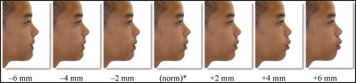
Sushner established African American lip position esthetic norms at 2 mm in front of the Ricketts’ E-line for males and 1 mm in front of the E-line for females. Kokich et al found, when observing altered dental esthetics, that the average size of clinically detectable differences was approximately 2 mm. Therefore, we used 2 mm as the range in which the raters could detect a difference from the E-line. Within that range, the patient profile was considered esthetically acceptable. The lateral cephalometric x-rays were digitally traced and the image calibrated to a 1:1 ratio. Each profile image was then linked to the corresponding lateral cephalometric x-ray. The linked pretreatment photographic image was then manipulated to produce different lip positions in 2-mm increments. All changes were confined to the anteroposterior dimension, with no changes in the vertical dimension. The structures manipulated for modified lip position were soft-tissue points between subnasale and the mentolabial sulcus. All initial photographic images were modified relative to Ricketts’ E-line to produce lower lip positions at −4, −2, 0, +2 (norm), +4, +6, and +8 mm for males and at −5, −3, −1, +1 (norm), + 3, +5, and +7 mm for females ( Fig 1 ). The 7 photos of each patient were saved in the database on a password-protected computer. These images were then further modified to produce the silhouette images. Each silhouette image was produced by adjusting brightness and hue to produce a 2-tone silhouette image. There were 7 modified photos and 7 corresponding silhouette images of each patient, for a total of 280 images. Each image was given a unique identifying number so that it would not be linked to the patient information in the Dolphin database.
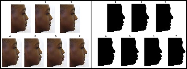
The original and the 6 modified photos of the same patient were placed together on a single slide so that there were 7 photos per slide for each of the 20 patients. The same was done with the silhouettes, yielding another 20 slides containing 7 silhouettes each. A presentation of the images was made with Microsoft Office PowerPoint Presentation software (Microsoft, Redmond, Wash). Each slide (of photos or silhouettes) was given a unique identifying number and then arranged haphazardly for presentation on a computer to the raters ( Fig 2 ), except that photos and silhouettes of the same patient were not presented consecutively.
The raters included 10 faculty orthodontists and 5 orthodontic residents with varying amounts of experience. Each rater entered his or her sex, whether a resident or faculty orthodontist, and the number of years experience on the data-collection form. All raters were white. There were 11 male and 4 female raters. Each rater was given a unique research number so that he or she could not be identified during the data analysis. Each was given an information sheet describing the research. At the beginning of the slide presentation, the raters received an explanation of the study and examples of the images that they were about to evaluate. Then each rater viewed the slide show, selected the numbered image (from among the 7 images) on each slide representing the most esthetic lip position, and indicated that number on the data form. The sex and age of each patient was placed at the top of each slide. Raters were given as much time as they required to complete the rating but were asked not to go back to slides they had already rated.
Statistical analysis
In the primary analysis, we compared the percentages of the photos and silhouettes preferred by the raters as most esthetic that were within 2 mm of the E-line (norm) in either direction. In 2 secondary analyses, we compared the percentages of the photos and silhouettes preferred by the raters that were flatter than the E-line (−4 or −6 mm) and fuller than the E-line (+4 or +6 mm) ( Table ).
| Distance from norm (mm) ∗ | Photos total (%) |
Photos male (%) |
Photos female (%) |
Silhouettes male (%) |
Silhouettes female (%) |
Silhouettes total (%) |
|---|---|---|---|---|---|---|
| −6 (flatter) | 0.33 | 0.67 | 0 | 15.33 | 1.33 | 8.33 |
| −4 (flatter) | 8.67 | 14 | 3.33 | 24 | 20.67 | 22.33 |
| −2 (acceptable) | 20 | 23.33 | 16.67 | 26.67 | 15.33 | 21 |
| 0 (at E-line/norm) | 39 | 36.67 | 41.33 | 22 | 44 | 33 |
| +2 (acceptable) | 26.67 | 22.67 | 30.67 | 9.33 | 14.67 | 12 |
| +4 (fuller) | 4 | 2 | 6 | 2.67 | 3.33 | 3 |
| +6 (fuller) | 1.33 | 0.67 | 2 | 0 | 0.67 | 0.33 |
∗ Male norm, +2 mm in front of the E-line; female norm, +1 mm in front of the E-line.
We used binary logistic regression with robust standard errors to account for the clustering (correlation) of the multiple ratings in each of the 20 patients. Some photos were presented more than once to the same rater. We used the Cohen kappa to assess intrarater reliability. The interrater reliabilities for photos and silhouettes were calculated as the average intraclass correlations (ICC) via a 2-way random effects models. All statistical analyses were performed with Stata (version 11; StataCorp, College Station, Tex) or SPSS (version 10; SPSS, Chicago, Ill) software.
Stay updated, free dental videos. Join our Telegram channel
VIDEdental - Online dental courses


