A girl, aged 7 years 11 months, came for treatment with the chief complaint of lack of eruption of her maxillary right central and lateral permanent incisors. The radiographic analysis indicated complete transposition of the maxillary right lateral incisor and canine, and a pseudotransposition of the maxillary right central and lateral incisors. This is a clinical condition of mixed transposition in the same hemi-arcade: the combination of a complete transposition and another incomplete or coronary transposition—a situation that has not been reported in the literature. The patient was treated with surgical exposure and ligation of the 3 teeth, the eruption was properly guided, and the correct order of the 3 teeth was restored in the arch. The diagnosis, appliance design, and treatment sequence are described.
Dental transposition can be defined as the positional interchange of 2 adjacent teeth, including their roots, or the development or eruption of a tooth in a position normally occupied by a nonadjacent tooth. A transposition is defined as complete when both the crowns and the roots are affected, or incomplete, also called pseudotransposition, when only the crowns are involved. It is a rare anomaly and a challenge for the clinician to diagnose and treat. Trauma is a commonly accepted etiologic factor in the development of a transposition, especially when the permutation involves a maxillary canine and a lateral incisor, or maxillary central and lateral incisors.
Because of the low prevalence and the variability, limited information is available about the treatment of patients with this disturbance. Therefore, it is a difficult task to standarize treatment procedures that could be extrapolated to similar clinical situations. Early identification and proper diagnosis are fundamental in adequate treatment planning and correction of these anomalies. In this case report, a unique situation of several transposed teeth in a hemi-arcade is presented, and the treatment sequence is described.
History and etiology
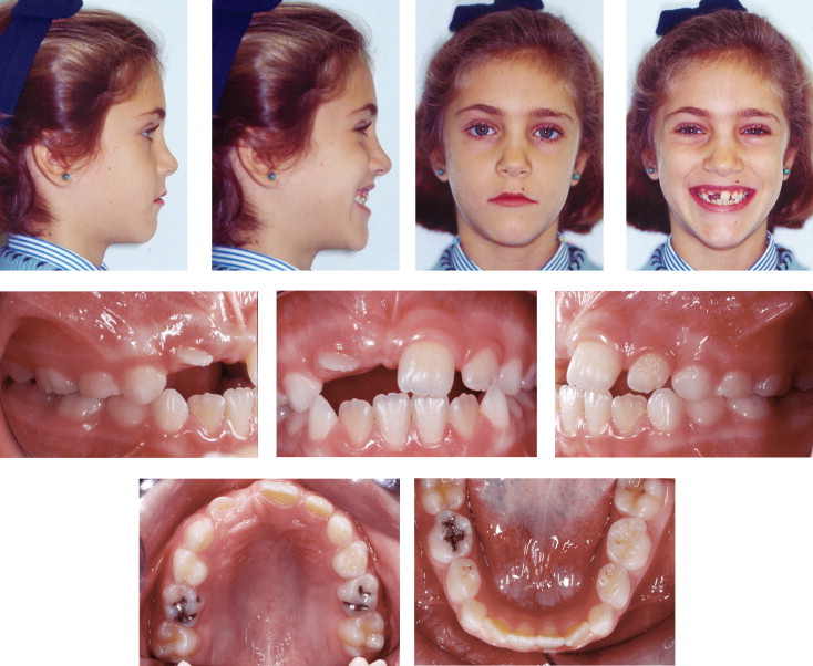
A girl, aged 7 years 11 months, came for treatment to our office in Seville, Spain, with the chief complaint of lack of eruption of the maxillary right central and lateral permanent incisors ( Figs 1–4 ). She had a history of a severe contusion to the anterior part of the maxillary dentoalveolus at the age of 1.5 years; it did not, however, result in apparent pulp damage or loss of teeth. She suffered another trauma to the same region when she was 4.5 years old, and this resulted in the loss of the maxillary right central and lateral deciduous incisors.

Diagnosis
The facial analysis showed an ovoid face, increased lower anterior facial height, and a mild increase of the gingival exposure on smiling ( Fig 1 ). Dentally, the patient exhibited a mild Class III molar relationship in the mixed dentition with a tendency toward an anterior open bite. The maxillary left central and lateral permanent incisors had erupted, but the maxillary right central and lateral permanent incisors had not ( Fig 1 ). In the area corresponding to the right central and lateral incisors, and in the area above the crown of the maxillary right deciduous canine, several prominences were identified by palpation of the mucosa that seemed to correspond to these unerupted teeth.
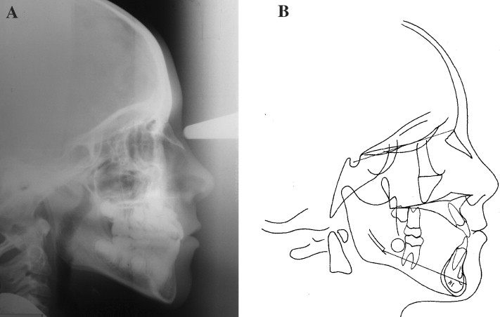
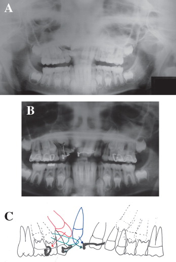
The cephalometric analysis showed a mild skeletal Class III malocclusion, with a growth axis that was predominantly vertical. There were an increase of lower anterior facial height and an open-bite tendency of both skeletal and dental origin ( Fig 3 , Table ). The panoramic radiograph showed 3 impacted teeth in the anterior region of the right side of the maxilla ( Fig 4 ): the right central and lateral permanenet incisors and the canine, in a triple mixed transposition. The crown of the lateral incisor was closer to the midline, whereas the crown of the central incisor occupied a more distal position. The canine was between the 2 incisors, positioned above them. The position of the central incisor was predominantly vertical, although its crown was oriented distally, causing an incomplete transposition with the crown of the lateral incisor. The lateral incisor had a horizontal angulation, with its crown toward the mesial aspect and its apex toward the distal apect. The incisal edge of the lateral incisor was visible through the gingiva ( Fig 2 ). The canine was positioned higher, with both its crown and root mesial to the lateral incisor, resulting in a complete transposition with the lateral incisor. The impaction of the 3 teeth was complete ( Fig 4 ).
| Value | Mean | Pretreatment | Postreatment phase 1 | Postreatment phase 2 | Retention |
|---|---|---|---|---|---|
| Sagittal relationships | |||||
| Maxillary position | |||||
| SNA (°) | 82±3.5 | 78 | 11 | 11 | 77 |
| Mandibular position | |||||
| SNPg (°) | 80±3.5 | 78 | 78 | 78 | 78 |
| Sagittal jaw relationship | |||||
| ANPg (°) | 2±2.5 | 0 | (−1) | (−1) | (−1) |
| Vertical relationships | |||||
| Maxillary inclination | |||||
| SN/ANS-PNS (°) | 8±3.0 | 6 | 8 | 10 | 10 |
| Mandibular inclination | |||||
| SN/GoGn (°) | 33±2.5 | 40 | 37 | 39 | 39 |
| ANS-PNS/Go Gn (°) | 25±6.0 | 34 | 29 | 29 | 29 |
| Dentobasal relationships | |||||
| Maxillary incisor inclination | |||||
| Maxillary incisor/ANS-PNS (°) | 110±6.0 | 109 | 109 | 110 | 111 |
| Mandibular incisor inclination | |||||
| Mandibular incisor/Go-Gn (°) | 94±7.0 | 88 | 90 | 83 | 84 |
| Mandibular incisor compensation | |||||
| Mandibular incisor/A-Pg (mm) | 2±2.0 | 4 | 4 | 3 | 4 |
| Dental relationships | |||||
| Overjet (mm) | 3.5±2.5 | 1 | 1 | 1 | 1 |
| Overbite (mm) | 2±2.5 | 1 | 1 | 1 | 1 |
| Interincisal (°) | 132±6.0 | 129 | 131 | 138 | 136 |
Diagnosis
The facial analysis showed an ovoid face, increased lower anterior facial height, and a mild increase of the gingival exposure on smiling ( Fig 1 ). Dentally, the patient exhibited a mild Class III molar relationship in the mixed dentition with a tendency toward an anterior open bite. The maxillary left central and lateral permanent incisors had erupted, but the maxillary right central and lateral permanent incisors had not ( Fig 1 ). In the area corresponding to the right central and lateral incisors, and in the area above the crown of the maxillary right deciduous canine, several prominences were identified by palpation of the mucosa that seemed to correspond to these unerupted teeth.
The cephalometric analysis showed a mild skeletal Class III malocclusion, with a growth axis that was predominantly vertical. There were an increase of lower anterior facial height and an open-bite tendency of both skeletal and dental origin ( Fig 3 , Table ). The panoramic radiograph showed 3 impacted teeth in the anterior region of the right side of the maxilla ( Fig 4 ): the right central and lateral permanenet incisors and the canine, in a triple mixed transposition. The crown of the lateral incisor was closer to the midline, whereas the crown of the central incisor occupied a more distal position. The canine was between the 2 incisors, positioned above them. The position of the central incisor was predominantly vertical, although its crown was oriented distally, causing an incomplete transposition with the crown of the lateral incisor. The lateral incisor had a horizontal angulation, with its crown toward the mesial aspect and its apex toward the distal apect. The incisal edge of the lateral incisor was visible through the gingiva ( Fig 2 ). The canine was positioned higher, with both its crown and root mesial to the lateral incisor, resulting in a complete transposition with the lateral incisor. The impaction of the 3 teeth was complete ( Fig 4 ).
| Value | Mean | Pretreatment | Postreatment phase 1 | Postreatment phase 2 | Retention |
|---|---|---|---|---|---|
| Sagittal relationships | |||||
| Maxillary position | |||||
| SNA (°) | 82±3.5 | 78 | 11 | 11 | 77 |
| Mandibular position | |||||
| SNPg (°) | 80±3.5 | 78 | 78 | 78 | 78 |
| Sagittal jaw relationship | |||||
| ANPg (°) | 2±2.5 | 0 | (−1) | (−1) | (−1) |
| Vertical relationships | |||||
| Maxillary inclination | |||||
| SN/ANS-PNS (°) | 8±3.0 | 6 | 8 | 10 | 10 |
| Mandibular inclination | |||||
| SN/GoGn (°) | 33±2.5 | 40 | 37 | 39 | 39 |
| ANS-PNS/Go Gn (°) | 25±6.0 | 34 | 29 | 29 | 29 |
| Dentobasal relationships | |||||
| Maxillary incisor inclination | |||||
| Maxillary incisor/ANS-PNS (°) | 110±6.0 | 109 | 109 | 110 | 111 |
| Mandibular incisor inclination | |||||
| Mandibular incisor/Go-Gn (°) | 94±7.0 | 88 | 90 | 83 | 84 |
| Mandibular incisor compensation | |||||
| Mandibular incisor/A-Pg (mm) | 2±2.0 | 4 | 4 | 3 | 4 |
| Dental relationships | |||||
| Overjet (mm) | 3.5±2.5 | 1 | 1 | 1 | 1 |
| Overbite (mm) | 2±2.5 | 1 | 1 | 1 | 1 |
| Interincisal (°) | 132±6.0 | 129 | 131 | 138 | 136 |
Stay updated, free dental videos. Join our Telegram channel
VIDEdental - Online dental courses


