Introduction
The purpose of this study was to investigate the changes of available mandibular space in the posterior dental arch of teenagers from 13 to 18 years old.
Methods
Longitudinal cephalograms of 28 adolescents (13 boys, 15 girls) with normal occlusion, selected from among 901 candidates, were taken annually from 13 to 18 years of age inclusively. Modified analyses with occlusal plane and occlusal plane perpendicular as reference planes were used to evaluate the changes of available space of the posterior mandibular arch.
Results
From 13 to 18 years of age, significant differences of mandibular posterior space were found among ages and sexes. The total increases of available space were 5.12 mm in the girls and 5.79 mm in the boys. For girls before age 16 and boys before age 17, the increased available space was contributed mainly by resorption of bone on the anterior border of the ramus. Mesial drift of the dental arch did not occur until the eruption of the third molars. The average available spaces increased 1.22 mm in girls less than age 16 and 1.45 mm in boys less than age 17 per side per year.
Conclusions
The prediction of available space in the posterior mandibular arch should be based on age and sex.
Total dentition space analysis is valuable for orthodontic diagnosis and design. The space analysis in the posterior dental arch is of great importance and can help orthodontists to achieve a consistently high-quality result. The posterior arch area should be included in a complete orthodontic treatment. It is meaningful to predict whether there will be sufficient space for the third molars to erupt in the early permanent dentition. It is imprudent to create a posterior discrepancy while making adjustments in other areas—the midarch or the anterior arch. It is also unadvisable not to use the excess space in the posterior area to alleviate midarch and anterior space deficiencies.
Contemporary treatment protocols involving molar distalizing mechanics and even arch expansion might be sound approaches for correcting some malocclusions. However, these techniques do not necessarily create space to accommodate all the teeth. Rather, they would seem to involve “borrowing” space for alignment, and, in some patients, this borrowed space might need to be paid back in other forms, such as other extractions after active treatment, less space available for the second molars, or impaction of the third molars. The third molars account for 98% of all impacted teeth.
Third molar impaction or eruption is important in clinical practice. The role of the mandibular third molars in late incisor crowding is controversial. Some studies indicated a small, but statistically significant relationship between third molar eruption and increased crowding of anterior teeth. Also, preservation of the third molars might be beneficial for orthodontic anchorage, prosthetic abutments, or transplants.
Factors that influence third molar eruption include skeletal growth pattern, direction of dentition eruption, dental extractions, root configurations, and maturation of the third molars. However, the most important factor seems to be the space available in the retromolar region. But sometimes the third molars have not erupted, and the development of the posterior dental arch is not complete, even after active orthodontic treatment.
If it could be determined more accurately at an early age that the third molars would erupt, then orthodontic treatment and prognosis could progress on a more positive basis, and the orthodontist would not need to be concerned with checkups of the patient until ages 20 to 22 years.
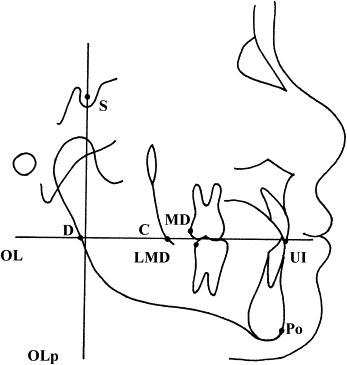
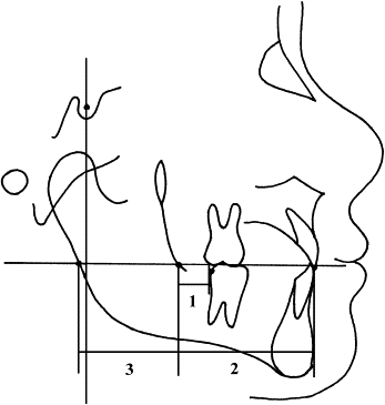
The aim of this study was to investigate the changes of posterior available space (distance between the distal contact point of the mandibular permanent first molar and the intersection of the occlusal plane with the anterior border of the ramus) in the mandible of adolescents from 13 to 18 years of age. This would provide important information for orthodontic clinicians.
Material and methods
We selected 75 subjects from 901 high school students. The selection criteria were (1) complete permanent dentition; (2) normal occlusion and normodivergent skeletal pattern with Class I canine and molar relationship (less than 3 mm of crowding in the anterior and midarch, less than 3 mm overjet, and overbite with less than one-third coverage of the mandibular incisor); (3) harmonious facial profile and competent lips at rest; and (4) no orthodontic treatment or dental extractions. These 75 adolescents agreed to participate in our research project, with cephalograms taken annually from 13 to 18 years of age inclusively. The study protocol was reviewed and approved by the institutional review board of Peking University. Because of the longitudinal nature and the aim of this investigation, subjects with less than 6 consecutive cephalometric observations were excluded. Thus, the investigation included 28 subjects (13 boys, 15 girls).
All cephalometric radiographs were taken with the same x-ray machine. Cephalometric landmarks were identified by 1 observer (L.-L.C.) under optimal conditions and then digitized with custom cephalometric analysis software.
When double projection caused 2 points or the right and left sides did not superimpose, the midpoint was used. The analysis of Bock and Pancherz with occlusal plane and occlusal plane perpendiculars as reference grids was used. The measuring points and reference lines used are shown in Figures 1 and 2 and defined in their legends.
Statistical analysis
The statistical analysis was performed with SPSS software (version 13.0, SPSS for Windows, SPSS, Chicago, Ill). The arithmetic mean and standard deviation were calculated for each variable. Analysis of variance (ANOVA) and paired t tests were performed. The level of significance was P >0.05.
Intraobserver reliability and reproducibility of the digitizer were checked on 20 randomly selected cephalometric radiographs that were retraced and redigitized 2 weeks later. The method error, S , was calculated as follows:
S x = ∑ D 2 2 N
Stay updated, free dental videos. Join our Telegram channel
VIDEdental - Online dental courses


