Fig. 11.1
Preoperative view of patient’s smile showing severe discoloration of the maxillary right canine (tooth #6, FDI 1.3) and generalized discoloration of the remaining dentition (A3, Vita Classical shade guide)
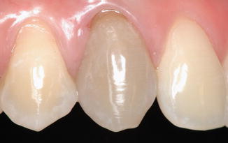
Fig. 11.2
Close-up magnification of tooth #6 (FDI 1.3)
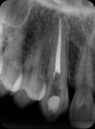
Fig. 11.3
Periapical radiograph of root-canal-treated tooth #6 (FDI 1.3)
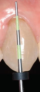
Fig. 11.4
A periodontal probe is used to determine the level of the epithelial attachment from the incisal edge of the tooth. This will serve as guide for placement of the root canal barrier (Chap. 8)
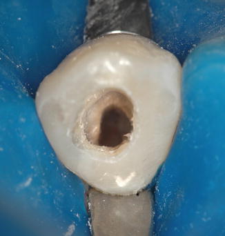
Fig. 11.5
After isolation with rubber dam and a #212 clamp, the restorative material was removed from endodontic access preparation. The root canal obturation material was removed from the cervical area
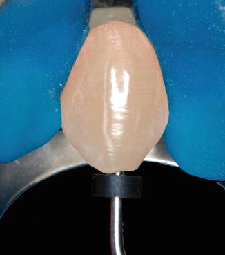
Fig. 11.6


The same periodontal probe is used to check if the root canal obturation material is removed to a level 2 mm apical to the CEJ, calibrated to the first measurement obtained with the periodontal probe as in Fig. 11.4
Stay updated, free dental videos. Join our Telegram channel
VIDEdental - Online dental courses
