Armamentarium
|
History of the Procedure
The goals of reconstruction in head and neck oncology may vary from patient to patient due to comorbidities involved with surgical reconstruction and patient motivation. Microvascular free tissue transfer has revolutionized the way surgeons address composite defects from ablative surgery of large tumors in a single-stage procedure. Furthermore, contemporary management of the patient with head and neck cancer integrates these surgical reconstructive techniques with prosthetic rehabilitation to optimize function and esthetics. The biology of the disease and the wound healing properties of the recipient site from prior therapy further affect the plan for reconstruction.
The complexity of rehabilitation for patients with maxillomandibular defects reconstructed with vascularized bone free flaps makes it necessary to devise treatment strategies that meet the patient’s expectations in terms of function, esthetics, and psychological and social aspects. Edentulous cancer patients who do not achieve oral rehabilitation after cancer surgery can exhibit significant psychological morbidity. Major advances in surgical reconstructive techniques and new approaches to maxillomandibular defects effectively provide a more conventional setting for prosthetic reconstruction of the dentoalveolar arch and surrounding structures.
Indications for the Use of the Procedure
Composite free flaps from the fibula, iliac crest, and scapula regions are designed and harvested to address tissue volumetric loss to restore mandibular continuity and to separate the oral cavity from sinonasal cavities. Soft tissue defects involving the overlying skin, mucosal defects involving the lip or cheek, and sensory and motor nerve deficits all define which reconstructive option is best for functional recovery. Preservation of tongue motion and the restoration of tongue volume are critical in obtaining a favorable functional outcome if tumor extension involves a significant portion of the tongue or the floor of the mouth. Vascularized bone–containing free flaps (VBFF) either restore continuity defects of the mandible or reproduce the stable base of the maxilla. Vascularized bone flaps from the fibula or iliac crest donor sites provide good to excellent bone volume and quality, which are required for osseointegration to enhance prosthetic rehabilitation. Composite free flaps from the scapula are selected when the soft tissue requirements of the defects are significant or when the use of the fibula donor site is contraindicated due to poor vascular runoff in the lower extremity or advanced age of the patient. However, this bone flap has a comparatively poor bone volume for osseointegration ( Table 24-1 ). If it is selected, two to four implants, no more than 10 mm in length, can be placed along the medial aspect of the angular tip for a removable prosthesis. Orientation of this area as a denture-bearing surface may prove challenging. Subsequent debulking of the overlying soft tissue most likely will be needed.
| Fibula * | Iliac Crest | Scapula | Radius | |
|---|---|---|---|---|
| Bone volume | ++++ | +++++ | +++ | + |
| Osseointegration | ++++ | ++++ | ++ | |
| Soft tissue | +++ | ++++ † | +++++ | +++ |
| Donor site morbidity | ++ | ++++ | ++ | + |
* The overall favorable characteristics of the fibular donor site makes this composite free flap the “workhorse” for jaw reconstruction compared to other donor sites.
The fact that a bone-containing free flap has its own blood supply offers additional strategies for implant-assisted prosthetic rehabilitation of acquired defects resulting from the treatment of large benign and malignant tumors of the jaws. One such strategy is to take advantage of the rich vascular bed for osseointegration before the delivery of adjuvant radiation therapy. Implant placement at the time of the initial reconstructive procedure also shortens the overall treatment time to a definitive prosthetic restoration. Once the bone has been fixed to the reconstruction plate and the anastomosis of recipient vessels is complete, implant placement can be performed before the insetting of the soft tissue used to restore the intraoral defect. After primary implant placement, the restorative team must allow 12 to 16 weeks for undisturbed healing and osseointegration of the fixtures. When the patient has completed radiation therapy after reconstruction and primary implant placement, the fixtures are uncovered, once the soft tissue reaction from the radiation treatment has subsided. At that time, soft tissue modification (e.g., flap debulking or vestibuloplasty procedures) can also be performed. A surgical stent can be used and secured to the implants for healing purposes before the fabrication of the definitive prosthesis. The surgical stent can be made with or without teeth, depending on the clinical situation and the patient’s wishes. The stent promotes undisturbed healing, maintains vestibular height, and improves the function and appearance of the lips and mouth.
Primary implant placement is the key to developing a comprehensive approach to ablative surgery, subsequent reconstruction, and prosthodontic rehabilitation with adjunctive radiation. This also holds true for implant placement into native bone at the time of tumor resection to optimize prosthetic rehabilitation without additional surgical reconstructive procedures. Primary implant placement can circumvent the need for hyperbaric oxygen before secondary placement of fixtures in patients who will receive radiation therapy after the reconstruction. In addition, primary implant placement minimizes time with an unstable prosthesis and compromised function in the edentulous patient. In 1998 the authors reported the success of implants placed into VBFF as the primary setting and subsequently irradiated as 86% ( n = 81 implants). This information was part of a patient cohort of 210 cases using microvascular composite flaps for oromandibular reconstruction.
Limitations and Contraindications
If a patient has received radiation therapy to the head and neck region, review of the simulation plan, including dosimetry and fields, is necessary to determine whether native bone or the VBFF has been affected, resulting in a compromised situation for osseointegration. Patients who undergo a hyperbaric oxygen protocol do so to enhance the vascularity of the surgical bed before implant surgery. Hyperbaric oxygen has been reported as beneficial to postirradiated native mandible and fibula free flaps.
The decision for fixed or removable prosthetic restorations depends on clinical factors such as bone availability, the number and position of implants to assist or support the restoration, and the hygiene maintenance and manual dexterity of the patient. In addition to these clinical factors, other considerations, such as comfort and psychosocial implications, affect prosthetic design. When addressing the reconstruction of the dental arch with osseointegrated fixtures, our preference is to provide patients with a fixed implant-supported restoration. When the remaining arch is edentulous and a lateral mandibular free flap reconstruction is performed, the fixtures should be placed in the anterior native mandible. This is an ideal location for implant placement in patients undergoing lateral jaw resection for posterior alveolar, floor of the mouth/lateral tongue, or tonsillar primary tumors because this area is usually spared radiation. A minimum of four or five implants, with the greatest anterior-posterior spread to minimize cantilever forces of the distal extension of the prosthesis, is recommended to restore the total dental arch. Posterior placement of the distal implant on the contralateral side of the mandible is potentially limited by the inferior alveolar neurovascular bundle and mental nerve. These landmarks must be identified, and care must be taken to avoid injuring them. Three or four implants placed into VBFF are recommended for unilateral maxillomandibular defects. As the defect crosses the midline, more implants are necessary to support the prosthesis; up to six implants are required to support a fixed restoration.
Issues regarding peri-implant soft tissue maintenance also arise. When muscle from the free flap is used for lining the oral cavity, the neomucosa around the implants might require repeated surgical débridement of hyperplastic inflammatory tissue around the transmucosal abutments. This problem may require excision and simple repair to possible split thickness skin grafting (STSG) if repeated robust growth of this unwanted tissue continues to be a problem.
Technique: Mandible Reconstruction
The placement of dental implants in a VBFF is not substantially different in principle or method from placement of implants in the native bone of the maxilla and mandible. The reader is encouraged to review Chapter 19 for technical information on the actual sequencing and implant placement details. Relevant differences in surgical technique and planning for implant placement in VBFF reconstructions of mandibular and maxillary defects are reviewed in the following sections.
Step 1:
Incision Design
For microvascular flaps with an intraoral skin paddle, the incision should be placed at the junction between the native oral mucosa and the skin of the free flap.
Step 2:
Flap Retraction
The dissection plane should be advanced to the level of the underlying bone. The surgeon should remain cognizant of the location of the perforating blood vessels to the free flap. A Doppler may be helpful in finding the location of these vessels.
Step 3:
Implant Placement
Implant placement is as previously described (see Chapter 19 ).
Step 4:
Closure and Soft Tissue Management
In cases of maxillofacial free flap reconstruction, secondary soft tissue procedures such as debulking or deepithelialization of the skin paddle often must be performed, along with implant placement, to facilitate a more favorable tissue emergence of the dental implants. These adjunctive procedures may need to be performed in advance of dental implant placement to obtain appropriate soft tissue contour.
The advantages of the fibula free flap have made this a “workhorse” flap for mandibular reconstruction of discontinuity defects. The length of the bone that can be harvested allows for near total mandibular reconstruction (from condyle to condyle). The low donor site morbidity and its distance for the primary surgical field allow for a two-team approach, which reduces overall surgical time and associated morbidity. Additionally, there is good to excellent bone stock for osseointegration. The bicortical nature of the fibula offers approximately 12 to 15 mm of bone height for endosteal implant placement. Unlike the scapula or iliac crest free flaps, which are monocortical in terms of implant fixation, implants placed into the fibula should engage both cortices to improve initial stability, osseointegration, and the ability to resist forces.
The fibula is tubular and triangular in cross section, and the three surfaces have dedicated characteristics. One surface has cutaneous perforators arising from the peroneal artery and vein; another surface is where the vascular pedicle runs; and the lateral aspect is used for internal fixation hardware that will secure the flap in position to allow for undisturbed healing of the osteotomized fibula and union to the remaining native mandibular stumps. Orientation of skin and will determine whether the base or apex of the triangle is oriented as the neoridge of the maxilla or mandible. These factors have significant implications for whether implants can be placed in the immediate setting at the time of the surgical reconstruction.
The use of fibula rather than iliac crest donor sites can present a geometric challenge for prosthetic reconstruction. As mentioned, the fibula is best positioned at the inferior border of the mandible to reproduce contours of the lower third of the face. This may lead to an intraoral height discrepancy with the native mandible. Additionally, because the alveolus is naturally positioned lingual to the inferior border, a neomandible created by placing bone at the inferior border can result in subsequent implants placed facial to the dentition in the opposing arch. In such cases, an implant-assisted, removable overdenture can be constructed so that lip and cheek support and oral competence are promoted. The use of a bar framework positioned lingual to implants can overcome the height discrepancy and facial position. The overdenture has small fenestrations at the base of the facial flange, thereby overcoming the facial position of the implants. The contours of the mandibular prosthesis can provide support to the lower lip to restore projection and symmetry to the lower face. The loss of motor function from injury of the marginal mandibular branch of the facial nerve can be ameliorated with this means of lip support.
The restoration of bilateral occlusal contacts, where occlusal guidance and protection schemes are restored to that of a fully dentate individual, is a critical step to optimal functional rehabilitation. The position of the mandible is determined both condylar by elements and by the dental occlusion. Reconstruction of the mandibular discontinuity defect with VBFF allows for both condylar determinants to function normally. The condition of the occlusion and dentition has an effect on function. The nonsensate nature of the VBFF reconstruction, other sensory deficits, and potential motor nerve to the lip and cheek make occlusal rehabilitation a restorable variable. A distinction is made between tissue-supported occlusal contacts and implant-supported tooth contacts and functional outcome. A further distinction is made between fixed and removable implant restorations. Rehabilitation with a fixed dental prosthesis (FDP) has better results for chewing capacity and esthetics and results in less physiologic discomfort and psychological disability than do removable prosthetic counterparts. The rigidity of an FDP and replacement of teeth with surrounding alveolus is a “near normal” design without the daily reminder of a removable prosthesis. These restorations are screw retained where the prosthesis is retrievable rather than a cemented restoration. This design consideration is an important factor in case direct visualization of tissue is necessary.
Treatment planning involves more implants rather than a minimum number for support of a fixed restoration. In the event of an implant failure, prosthetic success is still achievable with a shorter restoration of the dental arch or an implant-assisted overdenture, without added time or surgery.
Other approaches have been used to overcome this height discrepancy of fibula free flap reconstruction of the mandible. One is to position the fibula more superior and use the reconstruction plating system to reproduce contours of the inferior border. Another is the “double barrel” technique, in which the fibula is folded to increase the bone height and reduce the discrepancy between the occlusal plane and reconstruction.
Another surgical technique is distraction osteogenesis of the fibula to overcome the height discrepancy. One study reported a mean vertical bone height of 13.58 mm in five patients receiving 22 implants and dental restorations. However, infection of the distraction rod was a potential complication mentioned.
The authors recently reported a retrospective case series ( n = 28 patients) in which inclusion criteria for all patients were VBFF reconstruction and computer-assisted planning for FDP restoration in preparation for implant surgery. Implant success with immediate load (IL), provisional, and definitive FDP restorations in VBFF was reported for the first time in a patient cohort. Patients were evaluated for implant success, computed tomography (CT)-derived surgical templates, immediate provisional restorations, and prosthodontic framework design.
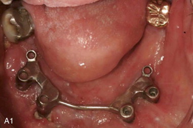
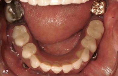
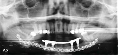
Ninety-nine of 116 osseointegrated implants placed were used for prosthetic restorations, achieving an 85.4% success rate. One hundred two implants achieved osseointegration (87.1%). Twenty-five of 28 patients (89.3%) received definitive implant FDP restorations. Two patients received removable implant-assisted restorations, and one patient was unable to complete rehabilitation due to implant failure. Thirteen of 28 patients received immediate or early-loaded fixed restorations at stage 1 implant surgery. The success rate for implants in the immediate restoration group was 89.3% (50/56). All 13 patients with immediate restorations had CT-derived surgical templates for their implant surgeries. Twelve of 13 immediate restorations were successful. Functional recovery for patients who undergo maxillomandibular reconstruction with VBFF is potentially more attainable with computer-assisted implant rehabilitation. Although the overall treatment time may be similar to that for patients without immediate restorations, we found the potential to provide patients with fixed provisional restorations during time for osseointegration and definitive FDP fabrication ( Figure 24-1 ).
Stay updated, free dental videos. Join our Telegram channel
VIDEdental - Online dental courses


