Growth and development

23

Growth and development

The processes by which a fertilized ovum develops finally into an adult human being are still not completely understood. Although measurements of hormone concentrations in blood and tissues are yielding more information about the stimuli for various developmental changes, there is virtually no information on how the time scale and sequence of development and growth are programmed and realised. The factors which govern organ size and shape are still poorly identified. Molecules known as organisers probably diffuse between cells to control differential growth and cell membranes probably interact with each other in delineating tissue boundaries – but no organiser molecules have yet been isolated and the mechanisms of membrane interaction are obscure.

Although the mechanisms are not known, it is possible to say that genetic factors play a major role in control. Homozygous twins reared under different conditions have similar body shapes even if actual size is different. The rate of physical development of a child, in the absence of major environmental differences, lies between those of its parents. Environment influences the extent to which the inherited potential is displayed. Some environmental factors may be particularly important at critical times in development and less so at other times. Thus malnutrition is most harmful in delaying growth and development during the first five years of life. However, the effect of malnutrition is to delay rather than to prevent, and if full nutrition can be restored the individuals may finally achieve normal development. On the other hand, tissues whose cells do not continue to divide after birth, such as nervous tissue, may not recover from malnutrition occurring during fetal life. In general, malnutrition affects size rather than shape.

Since the human race shows sexual dimorphism, development is affected by the presence or absence of the Y chromosome, females having two X chromosomes and males an X and a Y. The sex of the child is, therefore, governed by the chromosome carried by the spermatozoon. Even in abnormal karyotypes such as XXY, XXX, XXXY and XO, the presence of the Y chromosome is the deciding factor in the sex of the individual.

Measures of growth and development

The most obvious way to assess or express development is by age. However, chronological age is not necessarily related to either physical or mental development. The age of the fetus may be expressed either as post-menstrual, that is, dated from the last menstrual flow, or as post-conception. The latter is approximately two weeks later than the former. The term intra-uterine (i.u.) should mean post-conception and will be used in that sense here. Birth occurs at approximately 40 weeks post-menstrual or 36–38 weeks post-conception. Fetal age is often expressed chronologically when the assessment has been made dimensionally, since tables are available relating crown-rump lengths to the ages of average embryos. Birth is a clearly defined event in time and hence age can easily be expressed in relation to it. The next defined point in life is puberty, or the beginning of adolescence. In Europe it occurs in girls between the chronological ages of 8 and 13 years and in boys between 9.5 and 13.5 years. These are the ages at which enlargement of the sexual organs is first observed, an event which in girls precedes the first menstrual period, or menarche.

Size, or height, is not a particularly reliable indicator of development but is used. As mentioned above, it is one of the principal ways of determining the age, or developmental age, of the fetus. The crown-rump length or the crown-sole length can be measured on X-ray pictures, and ultrasonic scanning can be used to measure head size, crown-rump length and abdominal circumference.

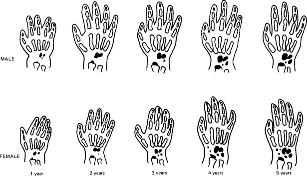
Other measures of development are available. The growth of the legs and feet is used as an indicator between birth and 18 months; after that the hand and wrist may be X-rayed to reveal the development of the individual bones which calcify in a specific sequence (Fig. 23.1). The mean ages at which the different stages are observed are termed bone ages. Growth in the female is always some 25% ahead of that in the male during the first ten years of life, and so female bone age is ahead of male bone age at the same chronological age.

Developmental age may also be assessed by the appearance of the teeth or, preferably, by X-rays of the teeth and jaws. The calcification and eruption of the deciduous teeth occur at similar rates in males and females, but calcification and eruption of the permanent teeth are earlier in females. The correlation between the age assessed from dental development and that assessed from the hand and wrist bones is not very strong, suggesting that the growth of these tissues is separately programmed and that there is not a close relationship between the development even of similar tissues.

The best correlation between the various measures of development is that between bone age and the age of the menarche in girls – but bone age does not correlate well with the age at which puberty begins in either sex.

Tissue growth

Tissues may grow by an increase in the number of cells, or an increase in the size of cells, or both. In normal development some tissues reach their definitive complement of cells early, and thereafter the number stays constant; other tissues are regenerative and have a continuing turnover of cells; whilst still others can show the property of regeneration only if the tissue is damaged or has to perform a much greater workload. Non-regenerative tissues include nerve, muscle and adipose tissue. Epithelia of the skin and the digestive tract are characterised by continuing death and replacement of cells; bone is constantly being laid down and resorbed. The liver and the kidneys are not normally subject to high rates of cell turnover but can show cell proliferation in response to increased metabolic load when large amounts of tissue are lost due to injury. An increase in organ size due to cell division is termed hyperplasia; an increase in organ size due only to increases in the size of cells is termed hypertrophy.

In the non-regenerative tissues growth occurs early in life. There are three phases: one of cell division, one of cell growth associated with a slower rate of cell division, and a final stage of cell growth only. Nerve cells stop dividing at about 18 weeks i.u. life and no new cell nuclei appear thereafter. The axons and dendrites can grow, and damage to one of these cell extensions is repaired by outward growth from the cell body. The supporting cells of the nervous system, the glial cells, appear to be capable of division throughout life. The growth and maintenance of sympathetic nerves is dependent upon nerve growth factor, a peptide produced in many tissues including the salivary glands, which is taken up by the axons and transported back to the soma, or body of the cell. Muscle cell numbers are also established at an early age, probably soon after 30 weeks i.u. life. Developing skeletal muscle is a tissue in which cells fuse together during embryonic development so that the final cells have many nuclei. Some cells remain as reserve, or satellite, cells: these are mononuclear with very little cytoplasm around the nucleus. Growth of muscle cells occurs by the synthesis of new protein to increase the contractile protein content of each cell. Muscle growth in response to the stimulus of increased usage involves incorporation of some satellite cell nuclei into the existing fibres in addition to the increase in protein content. Since growth occurs by increased protein synthesis, it is dependent upon the anabolic hormones, testosterone, adrenal androgens, and growth hormone. Insulin is also anabolic in its effects. The third tissue in which the number of cells is determined before birth is adipose tissue. Like the cells of skeletal muscle, fat cells increase the size of the tissue by increasing the volume of their cell contents: in this case by storing fat. Loss of stored fat from within the cells decreases the tissue size, just as decrease in the actin and myosin content of disused skeletal muscle leads to wasting of the muscle. It is probable that in adipose tissue, again as in muscle, there are less well-differentiated cells which will not develop into mature adipocytes unless excessive fat deposition occurs. Some writers dispute whether fat cells are non-regenerative and believe that new cells can be produced in these conditions. The adipose tissue of the fetus includes a relatively large proportion of brown adipose tissue, whose forming cells resemble those of the more usual white adipose tissue but whose mature cells differ from them both in appearance and function. All the non-regenerative tissues increase in size by hypertrophy.

Growth of the regenerative tissues may be continuous and responsive to tissue changes due to cell death or damage: there are, in addition, a number of polypeptide hormones which influence the growth of different tissues. These include epidermal growth factor, fibroblast growth factor and the somatomedins produced in response to circulating growth hormone.

Growth of bone and other hard tissues must be considered separately, since in these tissues the main bulk of the tissue is extracellular, and consists of inorganic salts which may be deposited and, except for dental enamel, removed by normal cell activity. Bone is formed in two ways: by deposition into a cartilage precursor, or by development in specialised fibrous tissue sites. After the initial formation, growth may occur either by cartilaginous growth followed by replacement with bone, or by remodelling as a result of activity of the cells of the periosteum in removing or building up layers of bone on the surface. The periosteum is a double layer of cells surrounding the bone; the outer layer is mainly fibroblasts, but the inner, or cambial, layer consists of cells which can lay down or resorb bone. In many bones, such as those of the limbs, cartilage cells form the shape of the fetal bones. The primary ossification centre appears in the centre of the shafts of these bones, and the cartilage is resorbed and replaced by bone. Shortly before birth, secondary centres of ossification form in the ends of the cartilaginous structure, and then bone is laid down in the articulating caps – the epiphyses. This leaves a plate of cartilage at each end of the bone; and it is in these plates that growth in length of the bone occurs. Control of such growth is largely exerted by somatomedin C produced in the liver in response to growth hormone. In addition there is control by testosterone, possibly the adrenal androgens, oestrogen and, in a permissive sense, by thyroid hormones. As growth progresses the epiphyseal plate of cartilage becomes thinner and finally the epiphyses fuse with the shaft, or diaphysis. This normally takes place towards the end of puberty, under the control of the gonadal hormones, the thyroid hormones, and growth hormone, acting in concert. Subsequent development depends upon the muscular forces acting on the bone which cause growth in response to muscle pull. Gravitational forces may supplement this. This remodelling includes bone deposition where there is mechanical stress, and resorption in its absence. The means by which stress is translated into growth is not yet known: it is possible that the piezo-electric forces generated between bone crystals may stimulate bone deposition. The final form of most bones, therefore, is one adapted to their function. The jaws differ from the limb bones in a number of ways. The mandible is not formed in a cartilaginous precursor, but develops from primary centres in fibrous tissue. The secondary centres are cartilaginous but only one of these, the articular cartilage, is near the end of the bone, and it is covered, not by a bony cap, but by a layer of fibrocartilage. This cartilage is not fully converted into bone until early adulthood – around 20 years. The jaws have a layer of bone in which the teeth are embedded, the alveolar process; this is appropriately placed to support the teeth, and its thickness and survival depend on the functioning of the teeth themselves. The whole of the alveolar process may disappear after the loss of the permanent teeth.

Only gold members can continue reading. Log In or Register to continue

Stay updated, free dental videos. Join our Telegram channel

Dec 5, 2015 | Posted by in General Dentistry | Comments Off on Growth and development

VIDEdental - Online dental courses

Get VIDEdental app for watching clinical videos