Almost one half of the patients who are admitted to a burn unit have some degree of facial burns. Most facial burns are superficial injuries typically caused by flash or splatter mechanisms. Extensive facial burns are uncommon and are usually the result of engulfing flame injuries from large explosions or house fires or scald injuries in children. In either case, the treatment of facial burns is different from that of burns in other locations. Facial tissue that is burned affects esthetic appearance and can negatively affect surrounding orifices such as the eyes, nose, mouth, and ears. Skin grafts are often a poor substitute for native facial skin, and specialized structures such as the eyelids or lips can never be completely restored to their original state.
The primary management of facial burns has a major effect on their outcome and is the subject of this chapter. Secondary burn reconstruction is a much larger topic that is not within the scope of this review.
Causes and Classification of Burns
The head, face, and neck are the anatomical sites most likely to sustain thermal injury. The most common cause of facial thermal injury is flash burns, and the least common cause is exposure to hot surfaces. The severity of facial burns depends on the duration of exposure to and the intensity of the burning agent (i.e., heat source). Time and temperature are the prime determinants of the severity of thermal injury; at temperatures between 44° C (110° F) and 51° C (124° F), the rate of cellular destruction doubles with each degree of increase in temperature. At temperatures higher than 51° C (124° F), exposure for 3 minutes or less can result in a full-thickness burn, and above 70° C (158° F), exposure for less than 2 seconds can lead to complete epidermal necrosis.
The specific heat source has an effect on the extent of the facial burn. Noncombustible hot liquids, such as water, are frequently encountered but usually at less than 70° C (158° F), and the duration of exposure is usually very short (<2 seconds) because the liquid runs off quickly from the body surface. Scald burns therefore typically result in only partial-thickness facial burns. Combustible hot liquids, such as grease, are usually heated to much higher temperatures (>100° C), and because of their viscous nature, the duration of exposure is much longer. This often creates contact areas of full-thickness burns. Flash burns to the face cause only a brief exposure to the heat source, resulting in a superficial or partial-thickness burn. The singed eyebrow or nasal hairs from flash burns may make the examiner suspect an inhalation injury, but the absence of carbonaceous soot or difficulty in breathing and the location of the injury make it unlikely that an inhalation injury has occurred. All of these patients should undergo bronchoscopic examination. Flame burns, particularly with ignition of clothing, lengthen exposure to intense heat and make a full-thickness injury more likely.
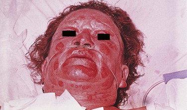
Burns are traditionally classified as first-, second-, or third-degree injuries, depending on the depth and completeness of dermal involvement. A better anatomical classification is that of superficial, partial-thickness (superficial and deep), and full-thickness burn injuries. All superficial and most partial-thickness burns maintain some dermal circulation, and they are usually capable of healing without excision or grafting. This can be clinically confirmed by the appearance of blisters, pain, and capillary refill. This is the most common type of facial burn because the face is often exposed to heat sources such as flash explosions rather than direct contact with flames ( Fig. 22-1 ). Withdrawal or turning away is a natural protective reaction that frequently spares the face from more severe thermal damage.
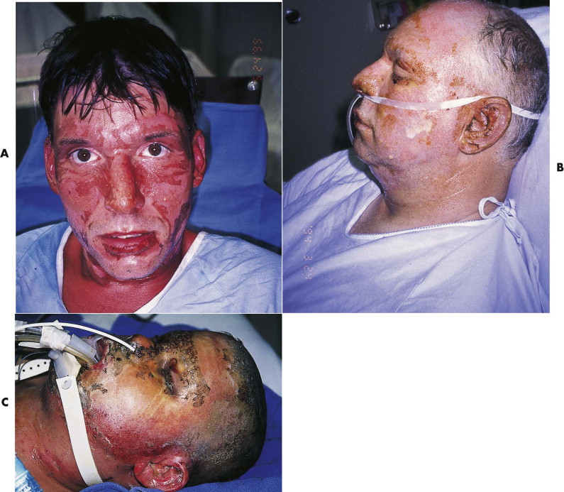
Full-thickness burns irreversibly damage the epidermis, papillary dermis, and reticular dermis and result in thrombosis of the subdermal plexus. Clinically, the skin appears pale, with minimal or no capillary refill. Hair shafts are removed easily. This degree of facial burn commonly results from prolonged contact with flames from bed or house fires when the victim is disabled by smoke, alcohol, or drugs ( Fig. 22-2 ). In extensive full-thickness burns, often called fourth-degree injuries, tissues and structures beneath the skin are involved. The skin is usually charred and insensitive. Causes include exposure to more than 1000 volts of electricity or to molten metal or prolonged contact with hot metal or flames. This degree of burn injury is rare in the face.
Describing facial burns as partial or full thickness provides a physiologically based classification system that also designates the approach to management. Although classifying the location and perceived depth of the initial burn is important, understanding the evolving nature of thermal injuries also is essential. It may take several days before the full extent and depth of a burn become apparent. Any early thoughts about surgical excision should be delayed until the burn depth and margins are clear. Interventions directed toward burn care, such as fluid resuscitation, antibiotics, and topical therapies, may help to prevent conversion of the burn to deeper levels.
Role of Topical Antimicrobials
The necrotic, nonviable tissue that covers the surface of a burn wound is a fertile medium for bacterial growth. Although excision of the burned tissue is the ultimate treatment for this problem, it is not done until demarcation of the burn is evident and the patient is adequately resuscitated and stabilized. In the interim, the use of topical antimicrobials is the mainstay of infection prevention. Although numerous bacteria, fungi, and viruses have been associated with primary infection of burn wounds, the most devastating are the gram-negative bacteria, particularly Pseudomonas . Infected burn wounds may easily progress in depth and extent. Intravenous antibiotics are often of little value because they cannot reach the site of inoculation due to vascular thrombosis.
The most commonly used topical agents are bacitracin, silver sulfadiazine, mafenide acetate, and collagenase ( Fig. 22-3 ). For superficial burns to the face, regular or ophthalmic bacitracin (Bacitracin, E. Fougera and Co., Melville, NY) is the preferred mode of treatment. It contains 500 mg/g of active agent and is a well-tolerated topical antimicrobial agent that causes minimal tissue reactivity. However, it offers no penetration of burn eschar, which is why its use is limited to superficial burns. In deeper partial-thickness and full-thickness burns, 1% silver sulfadiazine (Silvadene or Thermazene, Kendall Company, Marsfield, MA) is commonly used because it has the advantages of good penetration of burn eschar and painless application. It combines the antimicrobial action of silver ions with sulfadiazine. However, Silvadene tends to produce a pseudoeschar if not used appropriately, and this may confuse assessment of the depth of the burn. Mafenide acetate (Sulfamylon, Butek Pharmaceuticals, Morgantown, WV) at a concentration of 85 mg/g is a potent antimicrobial and has the best eschar penetration, but it is associated with pain after application. Silvadene and Sulfamylon must be kept away from the periorbital region because corneal ulceration, conjunctival edema and chemosis, and visual damage may result. In facial burns, all three antimicrobial agents are used in selective applications. In the periorbital area, bacitracin ophthalmic ointment is preferred, regardless of the depth of the burn, because it poses no risk to the eye. In the remainder of the face and scalp, 1% silver sulfadiazine may be liberally applied. Because of the known risk of cartilage exposure and suppurative chondritis from burns, Sulfamylon is used almost exclusively on the ear.
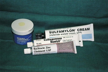
The use of an enzymatic débriding agent such as collagenase (Santyl Ointment, Knoll Pharmaceutical, Whippany, NJ) is helpful for superficial to deep partial-thickness burns. Santyl is a product of the bacterium Clostridium histolyticum. It can digest native and denatured collagen in devitalized tissue. In prospective studies, it improved healing and re-epithelialization times in partial-thickness burns compared with Silvadene. Because most of the dry weight of necrotic and viable tissue consists of collagen, a débriding collagenolytic enzyme seems to be an appropriate choice. It can chemically discriminate between viable and nonviable tissue, thereby preserving more viable tissue. This has particular value in the face, where every square millimeter of tissue has esthetic value. Antibiotic powders may be mixed with the ointment. Other débriding ointments include Accuzyme (Healthpoint Medical, San Antonio, TX), a papain-urea preparation (1,100,000 units of papain and 100 mg/g of urea). This débriding ointment is stronger than collagenase and produces significant pain on application. For this reason, I do not use it on the face.
State-of-the-Art Management
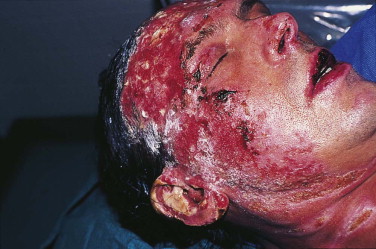
In the acute phase of facial burn management, it is important to rule out heat injury to the upper aerodigestive passages and lungs. The etiology of the burn injury is the single greatest determinant of inhalational injuries. Burns that have occurred outdoors or in open environments are unlikely to cause internal heat injuries. In closed environments, particularly house fires, internal injury should be suspected ( Fig. 22-4 ). Inhaled hot air can directly damage mucous membranes and cause edema. Swelling occurs within the first 24 to 48 hours and, if severe, may lead to airway obstruction and pulmonary insufficiency. In these cases, early endotracheal intubation should be performed. Nasotracheal intubation is preferred to the oral route because it can be easier to perform, is more comfortable when used for longer periods, and can be firmly secured. Fixation of the nasal tube is best done by transseptal suturing (2-3 or 3-0 silk), which provides good security and eliminates the need for tape on the face. When orally placed, the tube can be secured with interdental wires or sutures. If respiratory insufficiency is caused by heat injury, it is usually possible to extubate the patient after several days.
On admission, facial burns are débrided of all loose blisters and eschar. Although there is debate about whether to open blisters, leaving fluid-filled blisters on the face may be an impediment to wound evaluation and healing. In very superficial burns, particularly those involving the periorbital area, bacitracin ophthalmic ointment is used without dressings. Deeper burns may require more aggressive treatment, which is influenced by the anatomical location of the facial burn.
Tetanus prophylaxis is given to all burn patients unless a booster has been received in the past 5 years. Because routine administration of antibiotics does not protect against burn wound cellulitis or sepsis, their use in the acute setting is not recommended. Unless specifically indicated by obvious wound infection or positive culture results, liberal use of antibiotics contributes to the development of resistant organisms, a common problem in hospital burn units.
Scalp
Stay updated, free dental videos. Join our Telegram channel
VIDEdental - Online dental courses


