Introduction
This study was conducted to evaluate the effect of applying early orthodontic force on the shear bond strength (SBS) of orthodontic brackets bonded with 4 adhesive systems.
Methods
Eighty stainless steel brackets were bonded to the enamel surfaces of extracted premolars with 4 adhesive systems. For each adhesive, 10 brackets were bonded without application of force (groups 1, 3, 5, and 7), and another 10 were subjected to a 120-g force with a coil spring (groups 2, 4, 6, and 8). This force was applied 30 minutes after bonding and maintained for 24 hours. Groups 1 and 2 had Rely-a-bond primer and Rely-a-bond adhesive (Reliance Orthodontic Products, Itasca, Ill). Groups 3 and 4 had Transbond XT primer and Transbond XT adhesive (3M Unitek, Monrovia, Calif). Groups 5 and 6 had Transbond Plus Self Etching Primer and Transbond XT adhesive (3M Unitek). Groups 7 and 8 had RelyX Unicem (3M ESPE, Seefeld, Germany). After thermocycling, SBS testing was performed by using a universal testing machine (Type 500, Lloyd Instruments Ltd, Fareham Hants, UK). The results of SBS testing for all adhesives were analyzed by 2-way analysis of variance and the Duncan test. The unpaired Student t test was used to compare the effect of force on the SBS of each adhesive.
Results
Transbond XT primer and its adhesive had the highest values (without force, 11.2 ± 3.1 MPa; with force, 10.7 ± 2.7 MPa), and RelyX Unicem had the lowest (without force, 5.8 ± 1.5MPa; with force, 5.7 ± 1.6 MPa). Application of force yielded nonsignificant reductions in SBS for all adhesives; this reduction was less pronounced with RelyX Unicem.
Conclusions
For all studied adhesive systems, orthodontic force up to 120 g can be applied within the first hour after bonding with no deleterious effects on bond strength.
Since 1970, bonding of orthodontic brackets to tooth enamel has become an accepted clinical technique. A typical bonding procedure is based on alteration of the enamel surface by acid etching followed by application of adhesive primer and resin. The reduction of the number of steps for bonding procedures, reducing harm to the enamel surface, and minimizing bond failures during orthodontic treatment are of important clinical concerns. Self-etching primers (SEPs) were introduced to reduce clinical bonding steps and chair time because they combine the etching and priming steps. In addition, the use of acidic primers decreases the amount of residual adhesive on the enamel surface after debonding. Currently, a 1-step adhesive system has been introduced and used in restorative dentistry. It combines etchant, primer, and adhesive resin in 1 paste. It has several advantages, including a decrease in the possibility of contamination during bonding procedures and save chair time.
Many factors can affect bond strength between tooth enamel and orthodontic brackets, including type, composition and mode of curing of the adhesive, etching time and concentration of the etchant, bracket material and base design, loading mode, and oral environment. In addition, polymerization shrinkage and degree of conversion of orthodontic adhesives have a pronounced effect on the durability of bonding. In general, data collected after 24 hours have been used to determine the bond strength of orthodontic adhesives. However, materials used in the oral environment should be strong enough to withstand both short- and long-term forces. Some studies evaluated the effect of different testing times on the shear bond strength (SBS) of orthodontic brackets with various adhesive systems. These studies reported that bond strength increased over a short period of time. Although bond strength of orthodontic adhesives increased over the storage time, their initial stable times differed.
In clinical orthodontic practice, bonding of the brackets and placement of archwires might be done in the same visit. Hence, force could be applied to the bracket within the first hour after bonding, and, regardless of the relatively low magnitude of this force, it could have an adverse effect on bond strength. It was reported that polymerization of the adhesive should quickly reach a minimum value to enable the adhesive to resist bonding failure when tying in the initial archwire. In 1997, Ireland and Sherriff studied the effect of the timing of archwire placement on SBS, both in vitro and in vivo using a no-mix adhesive system. They found that preloading of the brackets 2 weeks before testing had no significant effect on SBS. At the same time, no significant difference on SBS was observed in patients who had archwires fitted in the same visit as bracket placement and those who had archwire placement delayed for at least 1 week after bonding. Ching et al investigated the influence of early loading on both shear and tensile bond strengths of a no-mix orthodontic adhesive. A static load of 78 g was applied 15 minutes after bonding and maintained for 2 weeks. They reported that the applied load had no significant effect on either shear or tensile strength of the studied adhesive. The aim of our study was to evaluate the effect of applying a continuous orthodontic force for 24 hours (30 minutes after bonding) on the SBS of orthodontic brackets bonded with 4 adhesive systems.
Material and methods
Eighty freshly extracted maxillary first premolars were collected, cleaned, and stored in a 0.1% aqueous thymol solution. The teeth selected had no cracks, caries, attrition, or restorations. They were embedded in autopolymerizing acrylic resin (Duracryl, SpofaDental, Prague, Czech Republic) poured in plastic rings with the buccal surface up. The teeth were cleaned and then polished with pumice and rubber cups. A hook made of 0.9-mm stainless steel round wire was fixed in the acrylic toward the apex of the teeth. Standard twin edgewise metal orthodontic brackets (American Orthodontics, Sheboygan, Wis) were used in this study with an average base area of 0.0184 in 2 (11.85 mm 2 ). The teeth were divided randomly into 8 equal groups of 10 premolars each. The brackets were bonded to the teeth by using 1 of 4 adhesive systems according to the manufacturers’ instructions. These adhesive systems are described in Table I .
| Adhesive system | Description | Manufacturer | Batch number |
|---|---|---|---|
| Rely-a-bond primer, Rely-a-bond adhesive | Chemical-cured, total etch, 2-component system | Reliance Orthodontic Products, Itasca, Ill | 0706277, primer 0707032, adhesive |
| Transbond XT primer, Transbond XT adhesive | Light-cured, total etch, 2-component system | 3M Unitek, Monrovia, Calif | 6EC, primer 6XL, adhesive |
| Transbond Plus SEP, Transbond XT adhesive | Light-cured SEP, 2-component adhesive system | 3M Unitek, Monrovia, Calif | D 4775 261899, SEP 6XL, adhesive |
| RelyX Unicem | Dual-cured, self-etching, self-priming, 1-component system | 3M ESPE, Seefeld, Germany | 286940 |
In groups 1 and 2, the enamel surfaces were etched with 37% phosphoric acid gel (Total Etch, Ivoclar, Vivadent, Schaan, Liechtenstein) for 30 seconds, thoroughly rinsed with oil-free air-water spray for 15 seconds, and dried with oil-free compressed air. A coat of liquid Rely-a-bond primer (Reliance Orthodontic Products, Itasca, Ill) was applied on the etched enamel surface with a brush tip. Rely-a-bond adhesive (Reliance Orthodontic Products) was applied to the base of the bracket and pressed firmly onto the tooth. Excess adhesive was removed around the base of the bracket before setting. In groups 3 and 4, the etching, rinsing, and drying procedures were similar as in groups 1 and 2. A thin coat of Transbond XT primer (3M Unitek, Monrovia, Calif) was applied on the etched enamel. Transbond XT adhesive paste (3M Unitek) was applied to the base of the bracket and pressed firmly onto the tooth. Excess adhesive was removed around the base of the bracket, and the adhesive was light cured (Megalux, Mega-Physik Dental, Rastatt, Germany) on each interproximal side for 10 seconds.
In groups 5 and 6, the unconditioned enamel was treated with Transbond Plus SEP (3M Unitek). The contents of the package were mixed together, applied, and rubbed on the enamel surface for 3 to 5 seconds. A moisture-free air source was used to deliver a gentle burst of air to the primer. The surface was lightly air dried for 5 seconds. The bracket was bonded with the same bonding resin and curing light as used for groups 3 and 4.
In groups 7 and 8, the RelyX Unicem capsule was activated in the Aplicap Activator (3M ESPE, Seefeld, Germany); then the capsule was mixed for 15 seconds in a high-frequency mixing unit. The capsule was placed in the Aplicap Applier (3M ESPE), and the cement was applied to the base of the bracket, which was pressed firmly onto the tooth surface. Excess adhesive was removed around the base of the bracket, and the adhesive was light cured for 10 seconds on each interproximal side.
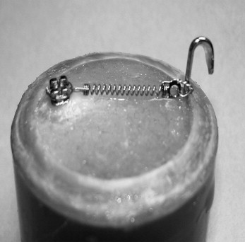
For all groups, the brackets were placed in their correct positions on the enamel surface and pressed with a compressive force of 300 g for 10 seconds by using a force gauge. After bonding, the specimens were allowed to bench set for 30 minutes. In groups 2, 4, 6, and 8, a 120-g force was applied to the bonded brackets with a closed titanium coil spring (American Orthodontics). One end of the coil spring was ligated to the bracket, and the other end was stretched and ligated to the metal hook ( Fig 1 ) until the desired force was reached according to the force gauge. After preparation, all specimens were stored in distilled water at 37°C ± 1°C for 24 hours. Before testing bond strength, all specimens were thermocycled 500 times between 2 water baths at 5°C and 55°C with a dwell time of 30 seconds in each bath.
SBS testing was carried out for all groups with a universal testing machine (Type 500, Lloyd Instruments Ltd, Fareham Hants, UK). The specimens were fixed horizontally in a specially designed steel base anchored to the fixed member of the testing machine. A knife-edged steel rod was fixed to the movable upper member of the machine. The specimens were subjected to a shear load at a crosshead speed of 2 mm per minute until failure. The load was applied under the incisal wings of each bonded bracket and parallel to the long axis of each mounted tooth. The load required to dislodge each bracket was recorded in newtons, and SBS was calculated in megapascals by dividing the load by the cross-sectional area of the bracket base.
After debonding, the teeth and brackets were examined under 10-times magnification. The amount of the adhesive on the enamel surface of the teeth was assessed by using the adhesive remnant index (ARI). The ARI has a range of 0 (no adhesive left on the enamel surface) to 3 (all adhesive left on the enamel surface). Less than 50% of the adhesive left on the enamel is scored 1; more than 50% of adhesive left on the enamel is scored 2.
Statistical analysis
Mean SBS values and standard deviations were calculated for all groups. The data obtained for all adhesive systems with and without application of force were subjected to 2-way analysis of variance (ANOVA) and the Duncan multiple range test to compare their mean values. Unpaired Student t tests were used to determine the significance of force on SBS for each adhesive system. The chi-square test was used to determine significant differences in the ARI scores between groups and for each adhesive with and without application of force. Significance for all statistical tests was predetermined at P <0.05.
Results
The results of the 2-way ANOVA are given in Table II . Mean SBS values, standard deviations, and the results of the Duncan and t tests for the adhesive systems with and without application of force are shown in Table III . A graphic presentation of these values is shown in Figure 2 .
| Source of variation | df | SS | MS | F | P value |
|---|---|---|---|---|---|
| Adhesive | 3 | 290.2391100 | 96.7463700 | 18.43 | <0.0001 |
| Applied force | 1 | 3.8106450 | 3.8106450 | 0.73 | 0.3970 |
| Interaction | 3 | 1.5696050 | 0.5232017 | 0.10 | 0.9599 |
| Error | 72 | 377.8594400 | 5.2480478 | ||
| Mean SBS and SD (MPa) | t test | ||||
|---|---|---|---|---|---|
| Without application of force | With application of force | t | P | Overall mean | |
| Rely-a-bond primer, Rely-a-bond adhesive | 8.8 ± 2.0 B | 7.9 ± 2.8 B | 0.80 | 0.435 | 8.3 B |
| Transbond XT primer, Transbond XT adhesive | 11.2 ± 3.1 A | 10. 7 ± 2.7 A | 0.32 | 0.753 | 11.0 A |
| Transbond Plus SEP, Transbond XT adhesive | 7.8 ± 2.6 CB | 7.5 ± 1.3 B | 0.08 | 0.938 | 7.7 B |
| RelyX Unicem | 5.8 ± 1.5 C | 5 .7 ± 1.6 C | 0.55 | 0.592 | 5.6 C |
| Overall mean | 8.4 | 8.0 | |||
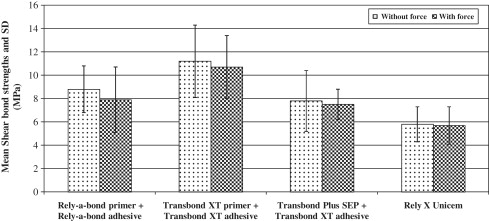
Regarding SBS without application of force, Transbond XT primer and its adhesive system had the highest value (11.2 ± 3.1 MPa), whereas RelyX Unicem showed the lowest value (5.8 ± 1.5MPa). Rely-a-bond primer and its adhesive system and Transbond Plus SEP and Transbond XT adhesive had intermediate values of 8.8 ± 2.0 and 7.8 ± 2.6 MPa, respectively. The results of 2-way ANOVA ( Table II ) indicated that, generally, there was a statistically significant difference ( P = 0.0001) in SBS among the 4 adhesive systems. There was no significant effect of the applied force on the SBS ( P = 0.3970). There was no significant interaction between applied force and type of adhesive ( P = 0.9599). The results of Duncan test ( Table III ) indicated a statistically significant difference between the SBS of Transbond XT primer and its adhesive system and the other adhesive systems. There was also a significant difference between Rely-a-bond primer and its adhesive system and RelyX Unicem ( P <0.05). On the other hand, there was no statistically significant difference between the SBS values of Rely-a-bond and Transbond Plus SEP systems and between RelyX Unicem and Transbond Plus SEP systems ( P >0.05).
With application of force, a slight reduction in the bond strength was shown for all adhesive systems. The results of the Duncan test indicated statistically significant differences between SBS values of Transbond XT primer and its adhesive system (10.7 ± 2.7 MPa) and the other adhesive systems, and between Rely-a-bond primer and its adhesive system (7.9 ± 2.8 MPa) and RelyX Unicem (5.7 ± 1.6 MPa). There was also a significant difference between Transbond Plus SEP and Transbond XT adhesive system (7.5 ± 1.3 MPa), and RelyX Unicem ( P <0.05). There was no statistically significant difference between SBS values of Rely-a-bond primer and its adhesive system and Transbond Plus SEP and Transbond XT adhesive system ( P >0.05).
When we compared the mean SBS values for each adhesive system with and without application of force, although all adhesive systems had a reduction in the SBS, the results of the unpaired Student t test showed no statistically significant differences ( P >0.05). The frequency distribution of ARI scores of the 4 adhesive systems with and without application of force is shown in Table IV . In general, failure was mostly cohesive with Rely-a-bond primer and its adhesive, and Transbond XT and its adhesive systems; it was mainly adhesive for Transbond Plus SEP and Transbond XT adhesive system, and RelyX Unicem. The results of the chi-square test indicated a statistically significant difference (22.41, P <0.05) in the ARI scores between the adhesives without application of force, but there was no statistically significant difference (15.63, P >0.05) between them with the application of force. For all adhesive systems, no statistically significant difference ( P >0.05) was obtained with and without application of force.


