Introduction
The purpose of the study was to examine the changes in stability of pharyngeal airway space (PAS) and hyoid bone position after 2 types of mandibular osteotomies in comparison with a control group.
Methods
The subjects included 46 Japanese women with skeletal Class III malocclusion. Twenty-five patients with mandibular prognathism underwent single-jaw surgery with bilateral sagittal split ramus osteotomy (SSRO), and 21 patients underwent bilateral intraoral vertical ramus osteotomy (IVRO). The control subjects included 30 volunteer women with normal occlusion. The treated subjects were assessed at the beginning of treatment, immediately after surgery, and after postsurgical treatment.
Results
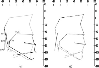
The Class III subjects had a significantly wider PAS than did the control subjects. Significant decreases in the lower PAS were observed after orthognathic surgery. The hyoid bone showed upward and forward movement with upward movement of the lower border of the PAS during the postsurgical stage in the SSRO group. In contrast, the anterior border of the PAS and the hyoid bone showed considerable backward movement in the IVRO group.
Conclusions
The posttreatment morphology of the PAS in both Class III groups approached that of the control group. The IVRO group showed a reduction in the airway dimensions, especially during the postsurgical period, which occurred during surgery in the SSRO group.
Editor’s comment
Mandibular setback surgery improves the occlusion, function, and esthetics by changing the position of the mandible, but it can also cause narrowing of the PAS and changes in the position of the hyoid bone and the tongue. This type of change is of concern because pharyngeal airway narrowing can cause obstructive sleep apnea. This study was designed to compare the changes of PAS and hyoid bone position between bilateral SSRO and bilateral IVRO after the release of intermaxillary fixation. The subjects consisted of 46 Japanese women who were diagnosed with mandibular prognathism and underwent single-jaw mandibular setback surgery with either SSRO (25 patients) or IVRO (21 patients) and orthodontic treatment.
The patients with Class III relationships had significantly larger PAS than those of the controls and showed significant posterior changes of the PAS immediately after surgery. The vertical position of the hyoid bone of the Class III patients moved remarkably downward and backward. Although the hyoid bone in the SSRO group returned toward its presurgical position after surgery, the hyoid bone in the IVRO group showed upward and backward movements during the same postsurgical period.
Although quite a bit has been written on this topic over the years, and 3-dimensional (3D) computed tomography evaluation of the upper airway is more state of the art, the lateral cephalometric evaluation that these authors used is highly correlated and has merits. Strengths of this study include the fact that only mandibular setback surgery patients were included; maxillary advancement and bimaxillary surgery patients (comprising most patients having Class III correction) were excluded. Other strengths included the control group, and that the subjects were women and well matched for age. On the other hand, the difference in the average amount of setback—8.1 mm for the SSRO group and 10.4 mm for the IVRO group—was rather large. In general, SSRO is used when substantial setback of the mandible is required, whereas IVRO is indicated for jaw deformity patients with unilateral temporomandibular disorders. Thus, the nature of mandibular setback is often different between SSRO and IVRO. Since soft-tissue adaptation is in a state of flux after treatment, a longer observation is needed to determine whether postsurgical morphology will sufficiently adapt to the improved lower facial morphology.