Introduction
The objective of this study was to compare the degrees of skeletal and dental asymmetry between subjects with Class II subdivision malocclusions and subjects with normal occlusions by using cone-beam computed tomography.
Methods
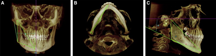
Thirty subjects with Angle Class II subdivision malocclusions (mean age, 13.99 years) and 30 subjects with normal occlusions (mean age, 14.32 years) were assessed with 3-dimensional cone-beam computed tomography scans. Independent t tests were used to compare orthogonal, linear, and angular measurements between sides and between groups.
Results
Total mandibular length and ramus height were shorter on the Class II side. Pogonion, menton, and the mandibular dental midline were deviated toward the Class II side. Gonion and the anterior condyle landmark were positioned more posteriorly on the Class II side. The mandibular dental landmarks were located more latero-postero-superiorly, and the maxillary dental landmarks more latero-antero-superiorly on the Class II side. There was loss of maxillary arch length, and the mandibular molar was closer to the ramus on the Class II side.
Conclusions
The etiology of Class II subdivision malocclusions is primarily due to an asymmetric mandible that is shorter and positioned posteriorly on the Class II side. A mesially positioned maxillary molar and a distally positioned mandibular molar on the Class II side are also minor contributing factors.
Editor’s comment
When I talk to experienced clinicians or board examiners, one of the most common complaints I hear is the misdiagnosis of and subsequent treatment problems younger orthodontists have with Class II subdivision malocclusions. Attempting to treat these patients without first diagnosing the source of the asymmetry often leads to long treatment times without resolution of the original problem. The objective of this study was to compare the degrees of skeletal and dental asymmetry between subjects with Angle Class II subdivision malocclusions and subjects with normal occlusions, based on true 3-dimensional (3D) analysis with cone-beam computed tomography (CBCT).
This is the first case-controlled study to use 3D volumetric imaging for the analysis of Class II subdivision malocclusions. Over 3000 patient records were reviewed to select the study sample based on specific inclusion and exclusion criteria. All sets of records included CBCT scans, dental models, and photographs. The most important factor initially considered was an occlusal assessment reflecting a Class II subdivision relationship. Evaluation of the other records confirmed the occlusal relationship found in the models; therefore, the Class II subdivision was consistent throughout the different records and thus most likely reflected the true occlusal relationship. It is important to highlight that a Class I occlusion does not mean a symmetric craniofacial complex. The importance of this factor is significant, since the intent of this study was to determine whether the Class II subdivision had a primarily dental or skeletal etiology. If the control sample had Class I occlusion but skeletal asymmetry (as could occur with dental compensation), analysis of the skeletal component would be significantly skewed because the “normal sample” was not really skeletally normal. Furthermore, the Class I occlusion subjects from this sample, who did not have apparent asymmetry, had some asymmetry when evaluated with a CBCT scan. This is why the authors believe that the between-sides evaluation might be more pertinent for the evaluation of symmetry than comparing the findings to a “normal group,” because mild asymmetries are common even in clinically symmetrical persons.
The primary contributing factor of a Class II subdivision malocclusion was a deficient mandible on the Class II side, which accounted for 61% of the total molar discrepancy between the groups. Of equal importance is the finding that there were no significant asymmetries among condylar pole measurements in the Class II subdivision malocclusion group. Significant dentoalveolar asymmetries were also present in the subdivision group.